14 May 2026

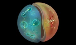
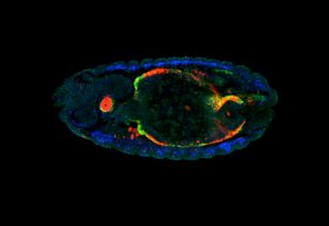
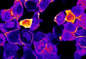
A newly identified protein, SNOR, has been found to help cells in a starvation-induced dormancy restart protein synthesis once nutrients are again available.
SCIENCE & TECHNOLOGY
16 December 2024
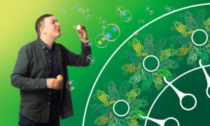
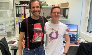
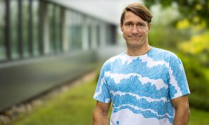
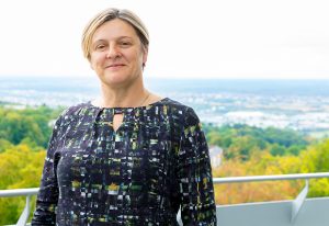
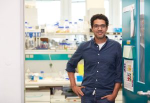
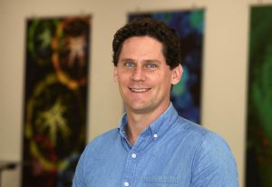
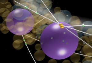
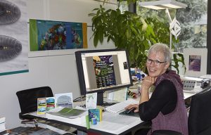
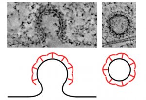
Veijo Salo, postdoc at EMBL Heidelberg, talks about seipin, the cell’s molecular ‘bubble blower’.
PEOPLE & PERSPECTIVES
8 October 2024
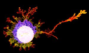
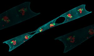
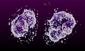
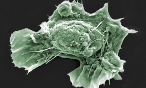
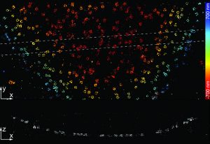
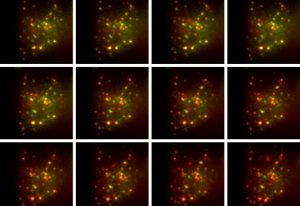
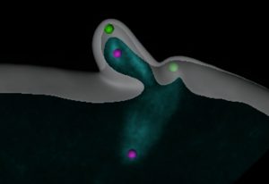
Scientists at EMBL Heidelberg and University of Virginia revealed a new cellular response to starvation: ribosomes attach to the mitochondrial outer membrane in a very unusual way, via their small subunit. The finding made in yeast might provide insights into how cancer cells survive the harsh…
SCIENCE & TECHNOLOGY
21 August 2024
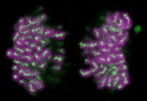
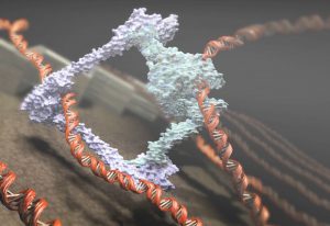
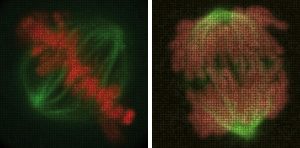
EMBL Heidelberg researchers discovered how a protein switches between repelling and gluing chromosomes during cell division. This helps the mother cell to divide the genome equally into two daughter cells and cluster chromosomes inside the daughter nuclei, ensuring a successful cell division.
SCIENCE & TECHNOLOGY
6 June 2024
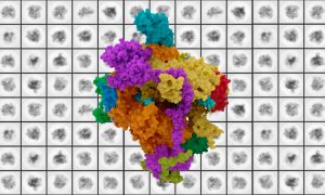
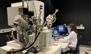
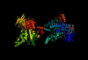
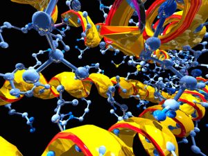
The group of Christian Löw at EMBL Hamburg and CSSB, and collaborators from the Christian-Albrechts-University Kiel and CNRS & Université Paris Cité worked together to reveal the structure and function of a previously unknown lysosome transporter, MFSD1.
SCIENCE & TECHNOLOGY
22 May 2024
New research by EMBL scientists shows how different modes of cell division used by animals and fungi might have evolved to support diverse life cycles.
SCIENCE & TECHNOLOGY
13 September 2023
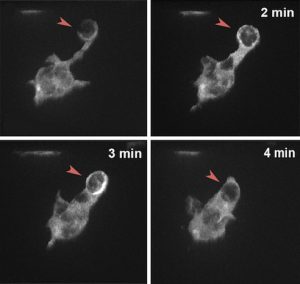
EMBL researchers have identified a novel mechanism that allows cells to sense obstacles in their path and avoid them while navigating complex environments.
SCIENCE & TECHNOLOGY
2023
sciencescience-technology
24 July 2023
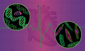
EMBL researchers have made new strides into understanding and reversing genetic defects that underlie familial heart disease.
SCIENCE & TECHNOLOGY
2023
sciencescience-technology
14 July 2023
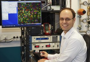
EMBL researchers use a new cell sorting technology to gain new insights into cellular function in health and disease, as well as for other innovative applications.
SCIENCE & TECHNOLOGY
2023
sciencescience-technology
5 May 2023
Dey Group holds second annual ‘labbatical’ to step outside daily research tasks with the help of single-celled model organisms.
LAB MATTERSSCIENCE & TECHNOLOGY
2023
lab-mattersscience-technology
31 August 2022
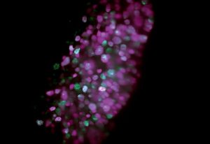
Physarum polycephalum, a single, giant cell containing tens of thousands of nuclei is large enough to be photographed with a phone.
SCIENCE & TECHNOLOGY
2022
picture-of-the-weekscience-technology
21 January 2022
EMBL researchers, in collaboration with BD Biosciences, have demonstrated a new technology that allows rapid image-based sorting of cells. The new technology represents a major upgrade to flow cytometry and has applications in diverse life science fields.
SCIENCE & TECHNOLOGY
2022
sciencescience-technology
9 December 2021
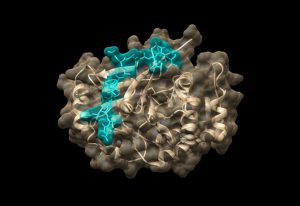
New structural biology research provides fundamental information critical to understanding enzyme mutations connected to rare diseases and cancers.
SCIENCE & TECHNOLOGY
2021
sciencescience-technology
29 November 2021

Using gene editing and three types of microscopy, one of EMBL’s newest group leaders is deciphering the functions of one of the smallest molecules involved in cell division, motility, and signalling, known as a centriole.
LAB MATTERSPEOPLE & PERSPECTIVES
2021
lab-matterspeople-perspectives
22 October 2021
A technology around since the ‘60s, flow cytometry has increasing applications. New leadership at EMBL’s flow cytometry facilities is looking to ease use, expand training, and encourage more collaboration.
LAB MATTERSSCIENCE & TECHNOLOGY
2021
lab-mattersscience-technology
12 October 2021
If researchers can identify specifically when good cells go bad, they can potentially understand disease better.
SCIENCE & TECHNOLOGY
2021
sciencescience-technology
5 October 2021
EMBL scientists and colleagues have developed an interactive atlas of the entire marine worm Platynereis dumerilii in its larval stage. The PlatyBrowser resource combines high-resolution gene expression data with volume electron microscopy images.
SCIENCE & TECHNOLOGY
2021
sciencescience-technology
30 August 2021
Giulia Zanetti from the Institute of Structural and Molecular Biology (ISMB) in London explains how the collaboration with the Cryo-Electron Microscopy Service Platform enabled her group to reveal the structure of protein transport complexes.
LAB MATTERSSCIENCE & TECHNOLOGY
2021
lab-mattersscience-technology
22 June 2021

Anna Erzberger, one of EMBL’s newest group leaders, will provide unique perspective as a theoretical biological physicist.
LAB MATTERSPEOPLE & PERSPECTIVES
2021
lab-matterspeople-perspectives
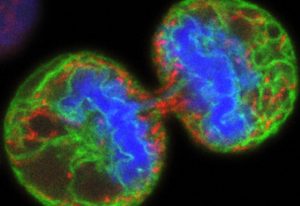
1 June 2021
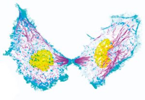
Captured by EMBL postdoc Arina Rybina, these ‘nuclear twins’ are two daughter nuclei straight after division of a HeLa cell.
SCIENCE & TECHNOLOGY
2021
picture-of-the-weekscience-technology
27 April 2021
EMBL scientists, together with collaborators from Heidelberg University, have provided further evidence of the gut’s role in COVID-19.
SCIENCE & TECHNOLOGY
2021
sciencescience-technology
27 April 2021
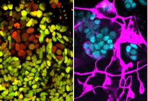
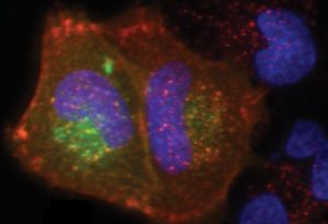
A page from a biologist’s colouring book? EMBL’s new interior wall design? Not quite – a bunch of liver cells, grown in the lab so that scientists can learn about fatty liver disease, or steatosis.
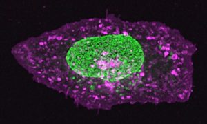
SCIENCE & TECHNOLOGY
2021
picture-of-the-weekscience-technology
5 February 2021
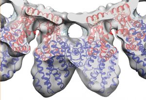
A new paper from the Galej group at EMBL Grenoble describes the structure of key parts of the Integrator complex, involved in gene expression.
SCIENCE & TECHNOLOGY
2021
sciencescience-technology
9 December 2020

Funding by the European Research Council (ERC) will support research on the timing of developmental processes in mammals
EMBL ANNOUNCEMENTSLAB MATTERS
2020
embl-announcementslab-matters
2 December 2020
Scientists in the Diz-Muñoz group at EMBL Heidelberg are working to build understanding of the role that mechanical properties play in affecting cell behaviour – a young and rapidly developing field of study. They have developed and successfully used a highly specialised technique to manipulate…
SCIENCE & TECHNOLOGY
2020
sciencescience-technology
27 October 2020
The nucleus of this cell fluoresces in bright green thanks to GFP-labelled nucleoporin proteins. EMBL scientists use engineered nucleoporins as 3D reference standards to improve super-resolution microscopy.
SCIENCE & TECHNOLOGY
2020
picture-of-the-weekscience-technology
18 September 2020
The internal clock that governs the development of embryos ticks slower for humans than for mice. Differences in the speed of biochemical reactions underlie the differences between species in the tempo of development.
SCIENCE & TECHNOLOGY
2020
sciencescience-technology
21 July 2020
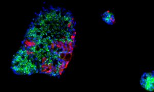
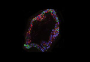
EMBL scientists have created a new, realistic 3D testbed that could help achieve the goal of stopping cancers before they start by studying cancer cells as they first form.
SCIENCE & TECHNOLOGY
2020
sciencescience-technology
30 April 2020
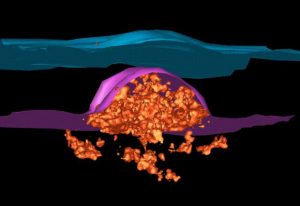
EMBL electron microscopy specialists collaborate with researchers from Heidelberg University Hospital to understand the changes occurring in cell structures upon SARS-CoV-2 infection.
SCIENCE & TECHNOLOGY
2020
sciencescience-technology
13 December 2019
Resource has implications for disease research
SCIENCE & TECHNOLOGY
2019
sciencescience-technology
18 November 2019
A new technique in cryo-EM
SCIENCE & TECHNOLOGY
2019
sciencescience-technology
11 March 2019

Scientists develop technology to measure how ATP concentration affects protein solubility in cells
SCIENCE & TECHNOLOGY
2019
sciencescience-technology
20 February 2019
How organs form in a mouse embryo
SCIENCE & TECHNOLOGY
2019
sciencescience-technology
9 October 2018
Meet Wendy Bickmore, Director of the MRC Human Genetics Unit, who spoke at the EMBL in the UK event
PEOPLE & PERSPECTIVES
2018
alumnipeople-perspectives
5 October 2018

Scientists in Finland met to share ideas and discover research opportunities
CONNECTIONS
26 July 2018
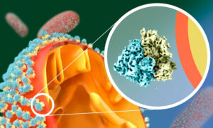
EMBL researchers visualise the proteins needed to capture molecules and bring them into a cell
SCIENCE & TECHNOLOGY
2018
sciencescience-technology
9 July 2018
New group leader at EMBL Barcelona creates artificial biological systems to study animal development
PEOPLE & PERSPECTIVES
2018
people-perspectivesscience
19 June 2018

More than 80 attendees gathered at the EMBL in Italy event at the FIRC Institute of Molecular Oncology (IFOM) in Milan
PEOPLE & PERSPECTIVES
2018
alumnipeople-perspectives
25 May 2018
EMBL alumna Melina Schuh recognised for excellence in science
PEOPLE & PERSPECTIVES
2018
alumnipeople-perspectives
9 April 2018
Open-source software allows standard microscopes to accurately image 3D structures
SCIENCE & TECHNOLOGY
2018
sciencescience-technology
9 April 2018
EMBL scientists count and locate chromosomal proteins during cell duplication
SCIENCE & TECHNOLOGY
2018
sciencescience-technology
20 March 2018
EMBL scientists discover how blood vessel cells become blood stem cells during embryonic development
SCIENCE & TECHNOLOGY
2018
sciencescience-technology
14 March 2018
Thanos Halazonetis discusses the EMBO/EMBL Symposium: DNA Replication: From Basic Biology to Disease
SCIENCE & TECHNOLOGY
2018
eventsscience-technology
16 February 2018

New EMBL group leader explores biophysical properties of chromosomes and other cellular assemblies
PEOPLE & PERSPECTIVES
2018
people-perspectivesscience
8 January 2018
New group leader studies sea anemones to investigate why some animals regenerate better than others
PEOPLE & PERSPECTIVES
2018
people-perspectivesscience
13 December 2017
New research reveals that two different mechanisms are responsible for chromosome folding
SCIENCE & TECHNOLOGY
2017
sciencescience-technology
7 December 2017

New research shows how pores form in the membrane that surrounds a cell’s nucleus
SCIENCE & TECHNOLOGY
2017
sciencescience-technology
23 November 2017
EMBL alumna Sigrid Reinsch trained as a cell biologist – now she helps run experiments in space
PEOPLE & PERSPECTIVES
2017
alumnipeople-perspectives
13 November 2017
A summary of recent research highlights from EMBL
SCIENCE & TECHNOLOGY
2017
sciencescience-technology
5 October 2017
EMBL researchers and collaborators unravel how chromosomes form
SCIENCE & TECHNOLOGY
2017
sciencescience-technology
11 August 2017
Meet Justin Crocker, EMBL’s new group leader in gene regulation during evolution and development
PEOPLE & PERSPECTIVES
2017
people-perspectivesscience
28 June 2017
Celebrating 40 years since the first EMBO electron microscopy training course
LAB MATTERS
14 June 2017
The Image Data Resource - prototype of the first open repository linking imaging and molecular data
SCIENCE & TECHNOLOGY
2017
sciencescience-technology
9 June 2017
EMBL researchers complete a molecular atlas showing gene expression in all cells in an entire animal
SCIENCE & TECHNOLOGY
2017
sciencescience-technology
6 June 2017
Two EMBL researchers are exploring new ways to filter out noise and get to the data they need
SCIENCE & TECHNOLOGY
2017
sciencescience-technology
15 May 2017
Hallmarks of residual breat cancer cells suggest new approaches for preventing relapse
SCIENCE & TECHNOLOGY
2017
sciencescience-technology
3 May 2017
ERC grantee Maja Köhn shares her vision for the next ten years
SCIENCE & TECHNOLOGY
2017
sciencescience-technology
27 April 2017
EMBL researchers develop an optical method for measuring the release of insulin from single cells
SCIENCE & TECHNOLOGY
2017
sciencescience-technology
11 April 2017
EMBL scientists add crucial knowledge to understanding of the bacterium that causes tuberculosis
SCIENCE & TECHNOLOGY
2017
sciencescience-technology
6 April 2017
Study by EMBL and DKFZ researchers means origins of myeloid leukaemias may need rethinking
SCIENCE & TECHNOLOGY
2017
sciencescience-technology
13 March 2017
ERC grantee Detlev Arendt shares his vision for the next ten years
SCIENCE & TECHNOLOGY
2017
sciencescience-technology
13 March 2017
ERC grantee Eileen Furlong shares her vision for the next ten years
SCIENCE & TECHNOLOGY
2017
sciencescience-technology
6 March 2017
EMBL researchers develop a computer model to explore the movement of nuclei in a multinuclear cell
SCIENCE & TECHNOLOGY
2017
sciencescience-technology
23 February 2017

Flies can do a lot for science, inside and outside the lab. EMBL alumna Isabel Palacios explains how
PEOPLE & PERSPECTIVES
2017
alumnipeople-perspectives
13 December 2016
Research on the effect of nerve cell stiffness on sensitivity to touch could lead to new painkillers
SCIENCE & TECHNOLOGY
2016
sciencescience-technology
7 December 2016
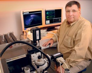
Paul Nurse’s failed experiment inspired a career that would uncover key mechanisms of cell division
SCIENCE & TECHNOLOGY
2016
sciencescience-technology
1 December 2016
EMBL’s Petra Riedinger retires after 40 years producing posters, graphics, artwork and more
LAB MATTERSPEOPLE & PERSPECTIVES
2016
lab-matterspeople-perspectives
24 November 2016

EMBL alumnus Jop Kind reflects on the questions that led him to this year’s John Kendrew Award
PEOPLE & PERSPECTIVES
2016
alumnipeople-perspectives
17 November 2016
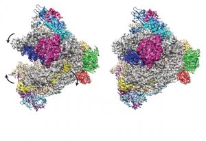
Cryo EM reconstruction of RNA Polymerase I reveals details of how molecule binds and transcribes DNA
SCIENCE & TECHNOLOGY
2016
sciencescience-technology
27 October 2016
Robert Prevedel develops deep-tissue microscopy for scientists to peer deep inside living organisms
PEOPLE & PERSPECTIVES
2016
people-perspectivesscience
23 September 2016
Puzzle of nuclear pore formation in growing nuclei solved
SCIENCE & TECHNOLOGY
2016
sciencescience-technology
1 August 2016
Molecular messengers synthesised to help study how cells respond to outside stimuli
SCIENCE & TECHNOLOGY
2016
sciencescience-technology
29 June 2016
New group leader combines physics and biology to answer the 'hows' of cell movement
PEOPLE & PERSPECTIVES
2016
people-perspectivesscience
14 June 2016
How cells eliminate protein deposits that can lead to neurodegenerative disorders
SCIENCE & TECHNOLOGY
2016
sciencescience-technology
20 May 2016

New method enables scientists to use light to direct where cancer cells go
SCIENCE & TECHNOLOGY
2016
sciencescience-technology
21 April 2016
How EMBL scientists are discovering and understanding the waves and rhythms inside us
SCIENCE & TECHNOLOGY
2016
sciencescience-technology
21 March 2016
1st real-time video of starfish egg cell eliminating crucial structures, to ensure embryo viability
SCIENCE & TECHNOLOGY
2016
sciencescience-technology
16 February 2016
How stem cells resist change
SCIENCE & TECHNOLOGY
2016
sciencescience-technology
17 December 2015
From initial development to a start-up company: Selective Plane Illumination Microscopy (SPIM) at EMBL.
SCIENCE & TECHNOLOGY
2015
sciencescience-technology
19 November 2015
Using lasers to shed light on how tissues get into shape
SCIENCE & TECHNOLOGY
2015
sciencescience-technology
30 October 2015
Fibres that pull membrane to form a vesicle exert a force that’s 2500 times a yeast cell’s own weight
SCIENCE & TECHNOLOGY
2015
sciencescience-technology
29 September 2015
Gold medal celebrates Eric Karsenti’s exceptional career and outstanding contributions to biology.
EMBL ANNOUNCEMENTSLAB MATTERS
2015
embl-announcementslab-matters
24 August 2015

Alumnus Thomas Vaccari reflects on the first joint symposium with EMBL Monterotondo, in Milan.
PEOPLE & PERSPECTIVES
2015
eventspeople-perspectives
20 August 2015
Collaboration between scientists reveals collaboration between lipids.
SCIENCE & TECHNOLOGY
2015
sciencescience-technology
10 July 2015
3D imaging unravels COPI coat of cells' transport vesicles.
SCIENCE & TECHNOLOGY
2015
sciencescience-technology
18 June 2015
Behaviour of clathrin proteins, crucial for endocytosis, is clarified using new imaging techniques.
SCIENCE & TECHNOLOGY
2015
sciencescience-technology
15 June 2015
Cells 'dance' as they draw together during early embryo development.
SCIENCE & TECHNOLOGY
2015
sciencescience-technology
21 May 2015

EMBL scientists demonstrate that spatial constraints are a key factor in determining nucleus size.
SCIENCE & TECHNOLOGY
2015
sciencescience-technology
5 May 2015
Stanford University biophysicist KC Huang on his collaboration with the Typas group in Heidelberg.
SCIENCE & TECHNOLOGY
2015
sciencescience-technology
20 April 2015
Ground-breaking microscopy technique gives unprecedented insight into endocytosis.
SCIENCE & TECHNOLOGY
2015
sciencescience-technology
16 March 2015
New fully automated technique enables scientists to chart complex protein networks in living cells.
SCIENCE & TECHNOLOGY
2015
sciencescience-technology
4 March 2015
Combining three different kinds of microscopy to determine how molecules move during endocytosis.
SCIENCE & TECHNOLOGY
2015
sciencescience-technology
25 February 2015
How strong does a spindle need to be? Videos put cell’s chromosome-separating machinery to the test
SCIENCE & TECHNOLOGY
2015
sciencescience-technology
4 February 2015
New microscopy-based method goes beyond gene sequencing, pinpointing the cause of disease.
SCIENCE & TECHNOLOGY
2015
sciencescience-technology
26 January 2015
EMBL scientists regularly receive prestigious awards – meet the latest honourees.
LAB MATTERS
18 December 2014

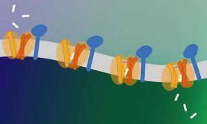
How do E.coli and similar bacteria grow safely? By using barrel-plugs as sensors.
SCIENCE & TECHNOLOGY
2014
sciencescience-technology
18 December 2014
Compound that can restore the function of poorly working mitochondria, with therapeutic potential.
SCIENCE & TECHNOLOGY
2014
sciencescience-technology
11 November 2014
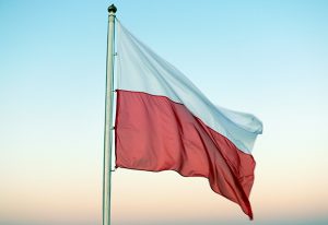
Alumna Anna Bartosik shares insights and hopes for EMBL's newest prospect member state, Poland.
LAB MATTERS
23 October 2014

Like sports teams, cells can huddle to communicate in secret and organise group behaviour
SCIENCE & TECHNOLOGY
2014
sciencescience-technology
20 October 2014
How Nobel-winning work by alumnus Stefan Hell shapes and inspires current EMBL scientists' research.
SCIENCE & TECHNOLOGY
2014
sciencescience-technology
17 October 2014
Flow cytometry: finding needles in haystacks
SCIENCE & TECHNOLOGY
2014
sciencescience-technology
12 September 2014
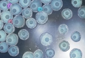
Researchers produce pristine stem cells, which can be precisely changed into clinically relevant cell types.
SCIENCE & TECHNOLOGY
2014
sciencescience-technology
21 August 2014
PhD Symposium poster reveals how a cell’s inner workings serve as both inspiration and toolkit.
LAB MATTERS
20 August 2014
Vasa protein preserves pieces of 'enemy' DNA to help protect the genes of future generations.
SCIENCE & TECHNOLOGY
2014
sciencescience-technology
17 July 2014
Cell biologists "underestimate the complexity" of protein interactions, says Toby Gibson.
SCIENCE & TECHNOLOGY
2014
sciencescience-technology
5 June 2014
A kaleidoscope of molecules is needed to clean up dead brain cells, and failure can have disastrous consequences
SCIENCE & TECHNOLOGY
2014
sciencescience-technology
18 May 2014
DNA-coralling protein complex in an unexpected bind
SCIENCE & TECHNOLOGY
2014
sciencescience-technology
9 May 2014
Genome Campus researchers discover that some immune cells turn themselves off by producing a steroid.
SCIENCE & TECHNOLOGY
2014
sciencescience-technology
1 April 2010
Name a human gene, and you’ll find a movie online showing you what happens to cells when it is switched off. This is the resource that researchers at the European Molecular Biology Laboratory (EMBL) in Heidelberg, Germany, and their collaborators in the Mitocheck consortium are making freely…
SCIENCE & TECHNOLOGY
2010
sciencescience-technology
4 March 2007
The European Molecular Biology Laboratory (EMBL) has developed a new computational tool that makes images obtained with cutting-edge microscopes even sharper. The technological advance and its applications are published in this week’s online issue of the journal Nature Methods. Since the…
SCIENCE & TECHNOLOGY
2007
sciencescience-technology
27 September 2006
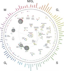
The life of a cell is all about growing and dividing at the right time. That is why the cell cycle is one of the most tightly regulated cellular processes. A control system with several layers adjusts when key components of the cell cycle machinery are produced, activated and degraded to make sure…
SCIENCE & TECHNOLOGY
2006
sciencescience-technology
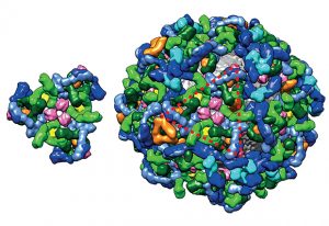
3 February 2005
Most things that happen in the cell are the work of ‘molecular machines’ – complexes of proteins that carry out important cellular functions. Until now, scientists didn’t have a clear idea of when proteins form these machines – are these complexes pre-fabricated or put…
SCIENCE & TECHNOLOGY
2005
sciencescience-technology
No results found