
Understanding early embryo development in mice
Single-cell sequencing helps researchers understand how mouse embryo cells generate different organs in the body
Researchers at the University of Cambridge and EMBL’s European Bioinformatics Institute (EMBL-EBI) have studied the genetic activity of over 100 000 embryonic cells to understand how different organs form in mouse embryos during early development. The research, published in Nature, provides information on how mammalian embryos develop during gastrulation, a key stage of development. This type of research could help researchers better understand the earliest stages of life.
Gastrulation is the process in animal and human development during which cells in the embryo become genetically programmed to generate all of the different organs in the body, including the brain, heart, lungs, gut and muscles. Despite its critical role in embryo development, scientists know very little about the full molecular profile of individual cells at this stage of life.
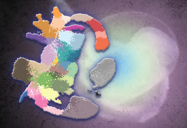
Interactive maps
The researchers used cutting-edge single-cell sequencing technology to measure the genetic activity in over 100 000 cells within the mouse embryo, between 6.5 and 8.5 days after fertilisation. Their results illustrate the great complexity of early development, while there are only a small number of distinct cell types at the start of gastrulation, within just 48 hours these cells branch out into over 30 different cell types with different genetic profiles.
Computational analyses enabled the scientists to generate interactive maps in which each cell is represented by a dot, and cells with similar molecular profiles are positioned close to each other. These new maps, which are freely available online, illustrate the trajectories of cellular development from one cell type to the next.
Mutant genes
The researchers tested the new molecular map by investigating a gene called Tal1. This gene is essential for normal blood development, but can also cause leukaemia if activated in the wrong cells. By comparing the reference atlas with over 10 000 Tal1 mutant cells, the researchers were able to decipher the consequences of the Tal1 mutation.
“By comparing our experimental data with the data collated within the molecular map, we were able to decipher precisely what was going on within the cells with mutant Tal1 genes,” explained John Marioni, group leader at EMBL-EBI and the Cancer Research UK Cambridge Institute. “We could see that the mutant cells weren’t simply getting stuck, or deciding to become a different cell type, but instead the cells started expressing a wide range of different genes, as if they were confused about what cell type they should mature into.”
“The map is also an invaluable reference point to understand how genetic mutations can disrupt embryo growth and cause developmental disorders and diseases,” said Berthold Göttgens, Professor of Molecular Haematology at the University of Cambridge.
This post was originally published on EMBL-EBI News.


