
From blood vessels to blood stem cells
EMBL scientists discover how blood vessel cells become blood stem cells during embryonic development
Cells – like those that make up our skin, heart, and brain – develop from stem cells. However, blood stem cells develop from vascular cells which line the walls of our blood vessels. EMBL Rome group leader, Christophe Lancrin, alongside first author Isabelle Bergiers, and their collaborators Tallulah Andrews and Martin Hemberg (Wellcome Sanger Institute), have published a paper in eLife focussing on the transcriptional regulation involved in this critical biological process.
What are the key findings of your paper?
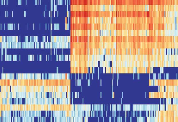
Our goal was to understand blood stem cell development during embryonic development. During this process, vascular cells change to become blood stem cells. We wanted to investigate which transcription factors (see fact box below) regulate this transition, and how. To do this, we looked specifically at a group of seven transcription factors which had already been described in blood cancers. By investigating the RNA in single cells using single cell transcriptomics, we found that cells in the middle of the transition between vascular and blood cell fates co-expressed all seven factors at the single-cell level.
This process can be modelled in the dish with embryonic stem cells (ESCs) differentiating into blood cells. We therefore engineered a special line of ESCs to simultaneously express these seven transcription factors in an inducible manner. This is the first time that so many transcription factors have been expressed in ESCs. What was very interesting was that the simultaneous expression of these transcription factors in the petri dish generated ‘undecided’ cells, expressing two identities at once (vascular and blood cell features), similarly to cells taken from the embryo which will go on to form blood stem cells.
Using advanced bioinformatics analysis, we predicted that some of these transcription factors had opposing activities. When we removed different transcription factors, from the original seven expressed in our engineered stem cells, we could induce either blood or vascular characteristics in the cell. Interestingly, we also saw that one transcription factor could have different functions, depending on which other regulators were present in these engineered cells.
Essentially, two cell fates are competing with each other in one cell. If one of the key transcription factors changes its role or is down-regulated, the transition to one cell type is accelerated. This could also be happening inside naturally-occurring ‘undecided’ cells lining the walls of our vessels. Knowing which transcription factors are important in determining either a blood or a vascular cell type is vital for us to further understand blood cell development.
What are the implications of this research?
Changes in cell fates also occur in diseases, like cancer. This occurs as part of the metastatic process when a cancerous cell becomes able to invade other tissues. Metastasis is a very well-researched topic but I don’t think anyone has studied it using the methods that we have used for studying blood cell development. It might be that this metastatic process is similar to what we describe in the current paper, with different transcription factors competing between two different cell fates. If we could better understand what the transcription factors are and how they compete with each other we could begin to think of ways to specifically inhibit this process and improve the chance of survival of cancer patients.

Gene Regulation
Transcription and translation are processes which occur within our cells. Together, they convert genes, encoded within our DNA, into functional proteins.
Transcription factors are proteins which bind to DNA to either activate or repress the transcription process of particular genes. During transcription, stretches of double-stranded DNA are separated to reveal DNA bases, the basic building blocks of DNA. Complementary RNA bases are added to the exposed DNA bases. The RNA bases are bound together in a strand and the RNA breaks free from the DNA.
The RNA can then be converted into a protein via translation. During this process, individual protein building blocks, known as amino acids, are encoded for by the RNA bases to form an ordered string of different amino acids. These 2D strings can be hundreds of amino acids long and are folded to create a 3D protein. Proteins have particular functions and this helps to determine the characteristics of the cell.


