Party at the nucleus?
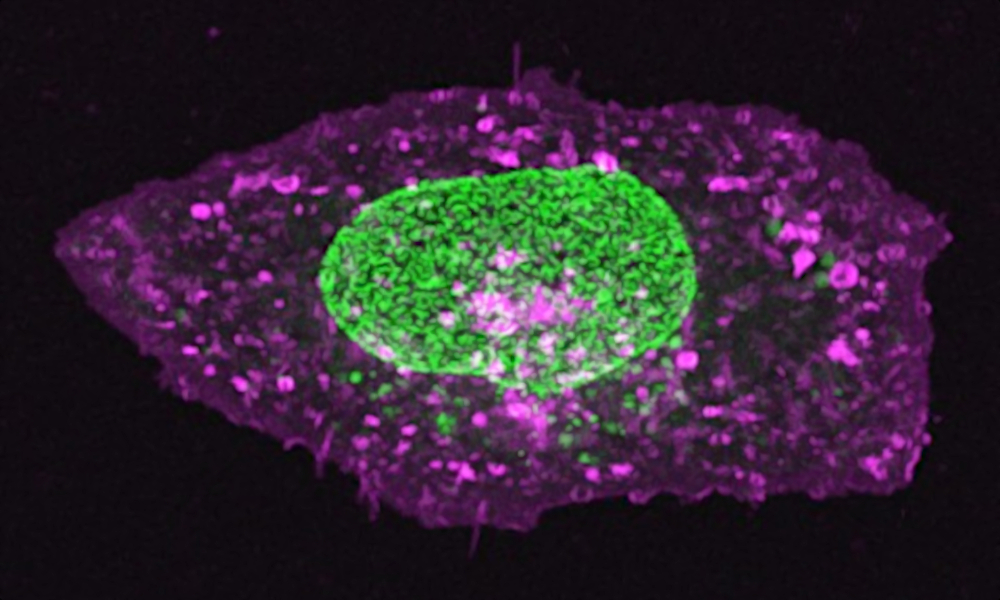
No, this is not an aerial view at a trendy night club, nor a new shade of fluorescent lipstick, or even a radioactive fried egg. Instead, this dazzler shows off a tool scientists have developed using nuclear pores as quality control standards for microscopy (shown in green).
Nuclear pores are protein-lined channels in the envelope that surrounds the nucleus, and they regulate the transport of molecules between the nucleus and the rest of the cell. Therefore, its structure has been studied in great detail. Building on this knowledge, the Ries group has engineered cell lines with nuclear pores labelled by different fluorescent proteins and dyes to act as three-dimensional ‘molecular rulers’. These can be used to precisely calibrate and optimize their microscopes. In this image, green fluorescent protein (GFP) labels the nuclear pore protein Nup96, and magenta shows a dye known as DiD, which highlights the membrane. This research showed how nuclear pores offer versatile reference standards that are widely shared with the research community in order to improve what researchers can learn from super-resolution microscopy.
Credit: Jervis Thevathasan & Markus Mund/EMBL
If you have a stunning picture of your science, your lab or your site, you can submit it here.