
The power of cell sorting: understanding heart disease, phase separation, plankton, and more
EMBL researchers are using a new cell sorting technology to gain new insights into cellular function in health and disease, as well as for other innovative applications.
The living world is made up of an astonishing variety of cells, and much of this diversity remains invisible unless one really goes looking. While identifying the characteristics of different cells has become easier and easier with advances in microscopy, scientists must also be able to isolate cells that look and behave differently from others to study them further.
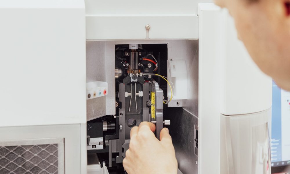
Since the 1970s, a technique called fluorescence-activated cell sorting (FACS) has helped make this possible, allowing researchers to isolate cells based on simple properties, like the presence of specific proteins on their surface or DNA content. Last year, in collaboration with BD Biosciences, EMBL scientists helped give FACS one of its biggest upgrades since its conception.
In a joint paper in 2022, EMBL researchers co-led by Lars Steinmetz, Sara Cuylen-Hãring, and collaborators at BD Biosciences and supported by the Flow Cytometry Core Facility described a method of capturing rapid-fire snapshots of cells and using these images to isolate cell populations based on complex characteristics, such as their shape and the specific location of proteins within cells. Capturing microscope-comparable images with such a level of detail at this very high speed had never been possible before.
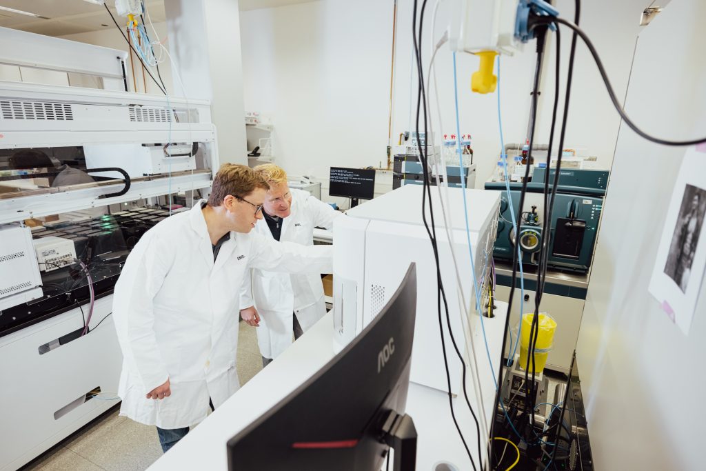
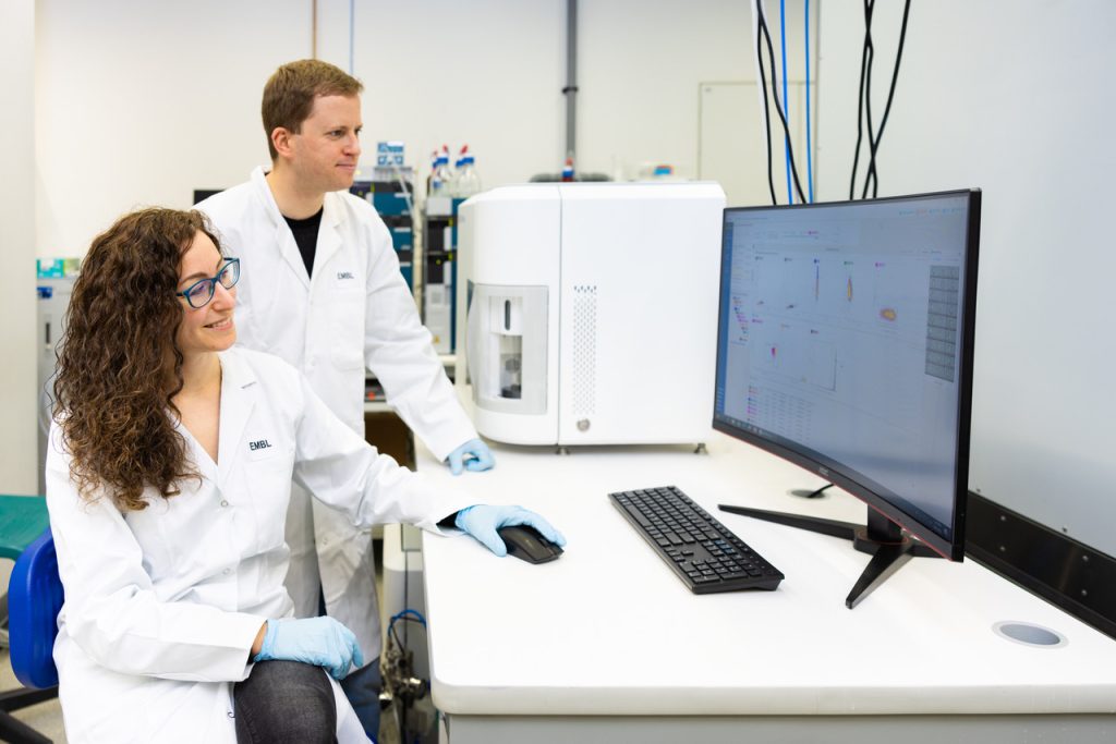
Since then, the technology, called image-enabled cell sorting (ICS), has been broadly adapted at EMBL, with support from the Flow Cytometry Core Facility, and used to expand the bounds of scientific knowledge. Scientists from the Steinmetz, Cuylen, and Pepperkok groups have deployed ICS to understand gene regulation in disease states, study phase separation in cells, and isolate novel plankton populations, among many other applications.
This video shows the functionality of image-enabled cell sorting. Credit: Sharmeen Ahmed/EMBL
“We’re now able to do experiments that we could only dream of before the development of ICS,” said Lars Steinmetz, Dieter Schwarz Foundation Professor and Associate Group Leader. “Our study in Science and ongoing work are examples of how massive leaps in technology, enabled by EMBL’s core facilities, open up completely new directions to accelerate our understanding of cell function, and what that means for health and disease.”
Probing the pathways of heart disease
For the Steinmetz group, the power of ICS lies in its high throughput. The increased scale of operations allows them to complete genetic screens that previously used to take weeks or months in a few hours or days instead. The team is currently using this method to dissect molecular pathways that contribute to the development of different types of diseases, such as heart diseases, one cell at a time and thousands of cells per second.
“Many diseases, such as cardiovascular and neurodegenerative diseases, involve proteins localising or aggregating where they shouldn’t be in the cell,” said Daniel Schraivogel, the lead author of the 2022 study and Research Staff Scientist in the Steinmetz Group. “ICS can help us figure out which cellular pathways are involved in driving protein mislocalisation during disease and to identify genes and drugs that can revert it.”
For this, the team is taking two approaches. The first incorporates giant mutation libraries of normal cells where specific genes have been selectively shut down. By testing for protein localisation, scientists can figure out which genes give rise to disease-like characteristics when lost. The second approach works in reverse: the team performs similar screens on cellular models of cardiovascular disorders to check whether the disruption of certain genes can revert a disease phenotype.
“Using these two approaches allows us to both understand the genetic basis for disease, as well as whether it can be reversed. This helps us identify additional drug targets and therapeutic approaches to treat cardiac diseases,” said Steinmetz.
Using ICS to understand phase separation
For the Cuylen Group, ICS helps with understanding fundamental cellular processes such as cell division or cellular organisation. For example, all cells have certain organelles that are not membrane-bound, such as the nucleolus. Recent studies have demonstrated that these membrane-less organelles assemble by liquid-liquid phase separation, similar to how oil and water separate from each other in solution. Given the fluid-like properties of these organelles as well as the cellular environment, it’s a mystery how these organelles maintain their structural organisation.
“It is an emerging field,” said Sara Cuylen-Häring, Group Leader. “We know very little about the regulators of phase separation. With this new technology, we can do unbiased genome-wide screens to see which molecules or pathways can regulate specific membrane-less organelles.” This would be difficult to achieve with traditional FACS, as the phenotypes being studied can only be analysed properly through imaging.
The Cuylen Group is also interested in cell division (mitosis). ICS allows scientists to isolate living cells in different stages of this process, which can subsequently be probed by various powerful analytical techniques such as proteomics.
“For the first time we can collect thousands of cells in very rare mitotic cell cycle stages.” said Cuylen-Häring. “The speed and specificity make it an ideal approach to investigate rare cellular events or phenotypes.”
Sorting plankton from the sea
Another equally exciting application of ICS is studying samples collected from the field. Hugo Berthelot, former postdoctoral fellow in the Pepperkok Team and current researcher at the French Research Institute for Exploitation of the Sea (Ifremer), is interested in plankton – tiny single-celled organisms that populate all of Earth’s oceans. Plankton come in a huge diversity of species, many of which have still not been identified or studied in the laboratory.
“We can use pigments and size to distinguish different populations of plankton,” said Berthelot. “ICS offers us a really cool way to simultaneously visualise and isolate these populations based on an unprecedented combination of factors.”
Marine plankton are one of the communities being sampled during the Traversing European Coastlines (TREC) expedition, EMBL’s Planetary Biology flagship project that kicked off in March 2023. With the advanced mobile services and laboratories, ICS may be a technology that soon transitions from the lab to the field.
“With this new and exciting technology, we now have a tool that allows us to use EMBL’s strengths in image analysis and multi-omic (single cell) analyses to develop new approaches and services that open completely new avenues for the characterisation of field samples at the molecular level,” said Rainer Pepperkok, EMBL’s Director of Scientific Core Facilities and Services.
Explorations continue
The development and application of ICS technology across scientific fields are central to an EMBL core mission – to actively engage in technology transfer and nurture industry-academia partnerships.
ICS also complements EMBL’s robust service offerings to the scientific community. “Adding imaging capabilities to cell sorting has opened up the possibility of identifying and correlating cell morphology and protein localisation with specific cellular processes,” said Diana Ordonez, Head of the EMBL Flow Cytometry Core Facility in Heidelberg. “The sentence: ‘Seeing is believing’ can finally be applied to the flow cytometry field!”
“We’re now starting to use ICS to understand the regulation of protein localisation and mislocalisation in a broad range of diseases. By combining ICS with other single-cell readouts, we can extract more information from each individual cell than ever before,” said Steinmetz. “This illustrates the importance of developing technologies and how they can advance our understanding of biology. ICS is a game changer for all life science disciplines, and we’ve only just scratched the surface of its applications.”


