
The hairy single-celled Tetrahymena provides a work reset
Dey Group holds second annual ‘labbatical’ to step outside daily research tasks with the help of single-celled model organisms
An ‘ordinary’ sabbatical might take one individual on a personal journey away from the daily grind to recharge batteries so they can return with a new professional perspective.
But why settle for the ordinary when you can do the extraordinary?
That could be the slogan for annual ‘labbaticals’ – two of which have now occurred in EMBL’s Dey group. The entire group halts day-to-day activities as much as possible to jointly pursue collective projects for two weeks. They apply their usual high-tech tools and evolutionary cell biology expertise, but with a totally different model organism and a goal of just seeing what happens.
Last year, the Dey group embarked on this research side step using Physarum polycephalum, an acellular slime mould. This year, Tetrahymena, a free-living ciliated microorganism, was the focal point. The goal was to have a ‘fun refresh’ and possibly jump-start scientific pursuits from this unusual exploration.
“The idea came from seeing how intensive sabbatical courses around the world have been known to lead to cool discoveries as scientists try a bunch of new, different things,” said Gautam Dey, EMBL Group Leader. “EMBL is a particularly good place for this since we have so many interesting tools and people are always willing to help. Here, if we dream it, we can probably do it.”
‘Slime mania’ for a refresh
Last spring, armed with a long list of experiment ideas, unbridled enthusiasm, and group ‘Slime Mania’ t-shirts made especially for the occasion, the Dey group looked to see what they could learn from Physarum, which is viewed as a sort of cellular computer, despite its blob-like, slime mould appearance.
“It was a very popular model system in the ‘60s and ‘70s, with a revival in recent years as people became interested in cellular computation,” Dey said. “There are some really cool experiments where scientists give Physarum multiple food sources on different parts of a plate, and the Physarum makes decisions about which one it prefers to eat, yet it’s just one cell.”
Despite its long-standing involvement in biological research, many unanswered basic cell biology questions about Physarum persist, such as how it divides and what triggers the switch between stages of its complex life cycle. New microscopy and other tools available at EMBL would likely help provide clearer answers.
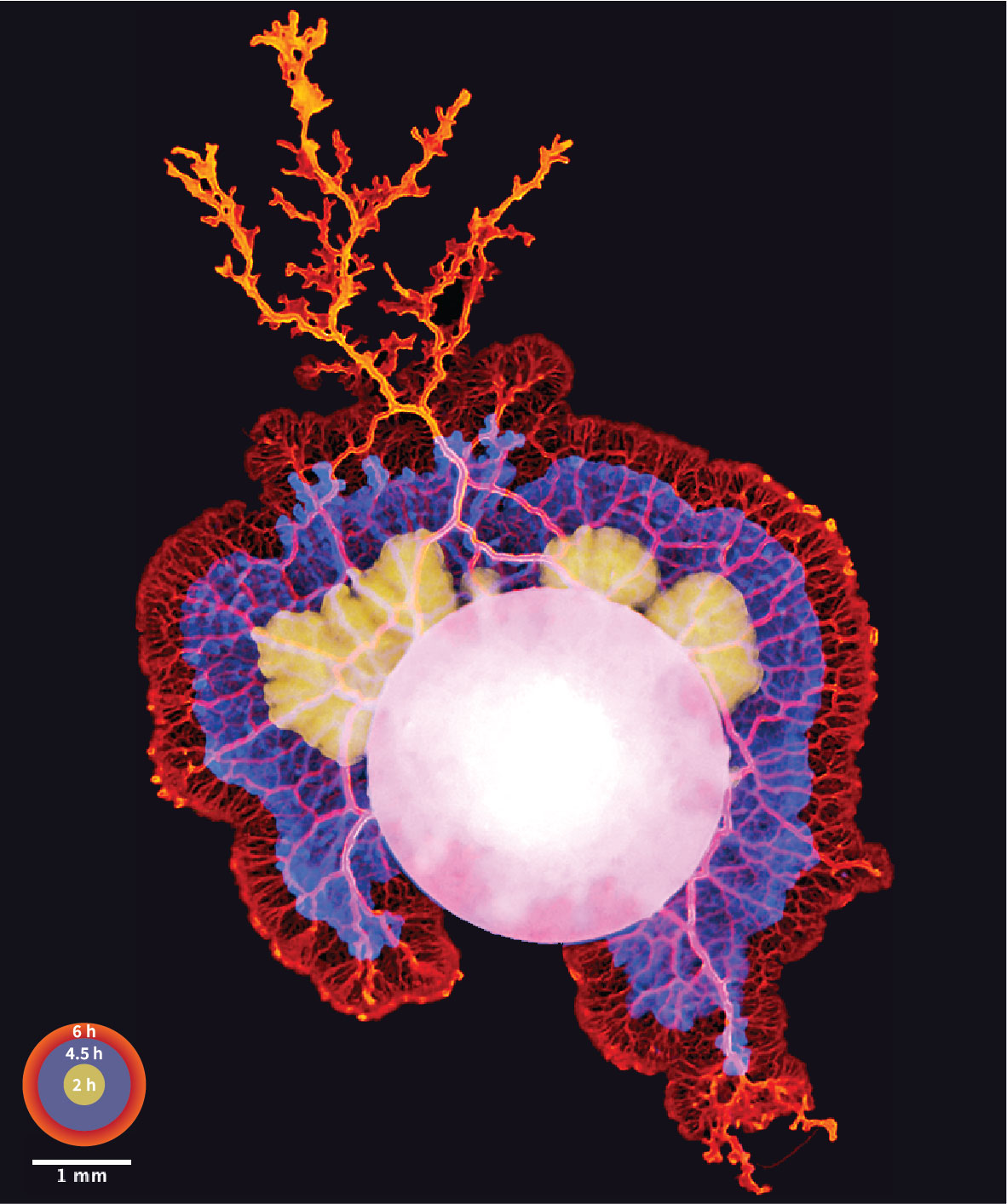
While the group only completed a small fraction of its intended experiments, the experience grew into two collaborative projects that new Dey group postdocs are now leading. It also prompted the team to link up with others interested in Physarum cell biology around the world, including the labs of Amy Gladfelter (Duke) and Karen Alim (TU Munich). Additionally, Felix Mikus, who was the group’s first PhD student to join in 2021, won the British Society for Cell Biology’s image competition in 2022 with his Physarum image taken without a microscope and with only a mobile phone.
Bring on the Tetrahymena
For this year’s ‘labbatical’, Tetrahymena seemed like a good choice. The Dey group is involved in EMBL’s TREC expedition doing field sampling along Europe’s coastlines for plankton, and many of their samples are full of ciliated organisms like Tetrahymena.
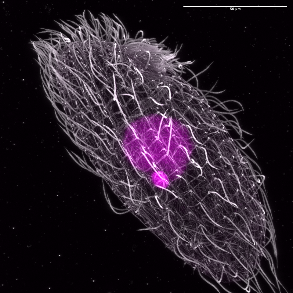
“Tetrahymena has very weird biology, but it’s everywhere,” Dey said. “They have many hair-like cilia on their body and use those for swimming, feeding, and so on. It’s also been responsible for two Nobel prizes.”
Tetrahymena were critical to discovering telomeres (the ends of chromosomes) and their role in ageing and replication. Interestingly, it has two nuclei. A tiny nucleus silently copies the genome and passes it on through sexual reproduction. Additionally, a mega nucleus contains many copies of the genome that it uses to grow and divide. The mega nucleus has thousands of mini chromosomes, so it has thousands of telomeres. That’s how Nobel Laureate Elizabeth Blackburn was able to make her finding. Additionally, Thomas Cech used Tetrahymena in his Nobel-recognised discovery that RNA can splice itself, possibly indicating that life might have started as RNA.
Tetrahymena’s long history as a model system meant it was easy for the Dey group to obtain the cells (in this case, a gift from Geert Kops at the University of Utrecht, and stocks ordered from the Tetrahymena Stock Center at Cornell University).
“This year, we prepared more ahead of time – and perhaps more realistically – but we still only managed to do about 15% of the experiments we planned,” Dey said. “We got some very nice images with a technique we’ve used a lot in the lab called expansion microscopy. While we know of other labs applying expansion microscopy to Tetrahymena, these studies are yet to be published.”
Mikus, in fact, will be one of the instructors in the inaugural edition of EMBL’s expansion microscopy course this summer.
“We tried some live imaging, which was very difficult because Tetrahymena swim very fast and they don’t like being immobilised, which we knew from the literature,” Mikus said. “They also don’t like being illuminated, which is not ideal for light microscopy.”
This year, with additional electron microscopy expertise from the Schwab team, the ‘labbatical’ discovered some features of Tetrahymena mitosis that had not been systematically reported in scientific literature.
Clearly, ‘labbaticals’ are catching on. The Dorrity group, which normally works with zebrafish to understand environmental impacts and responses at a cellular level, temporarily switched to sturgeon for their own recent group-wide break from day-to-day research.
“So now we have a little bit of expertise in this area, some of the tools, and some knowledge of what works and doesn’t,” Dey said. “In two weeks, you obviously can’t even get close to finishing a piece of science, but you can open many doors.”


