
Painting liver cells
Is this a page from a biologist’s colouring book? EMBL’s new interior wall design?
Not quite – what we’re seeing here is a bunch of liver cells, grown in the lab so that scientists can learn about the cellular processes involved in a disease known as fatty liver disease, or steatosis.
Steatosis is characterised by an excessive accumulation of fat molecules, or lipids, in our cells. The condition occurs primarily in the liver, where it can lead to liver cancer, but it can affect other organs as well. It’s often a result of obesity or excessive alcohol consumption, or is an accompanying symptom of other diseases, such as diabetes or hepatitis C.
In the lab, the processes leading to steatosis can be studied by growing liver cells in a dish containing a surplus of fatty acids – the building blocks of lipids. By adding other biomolecules that cells produce during disease or inflammation, scientists can cause the cells to take up more fatty acids than they need, mimicking steatosis.
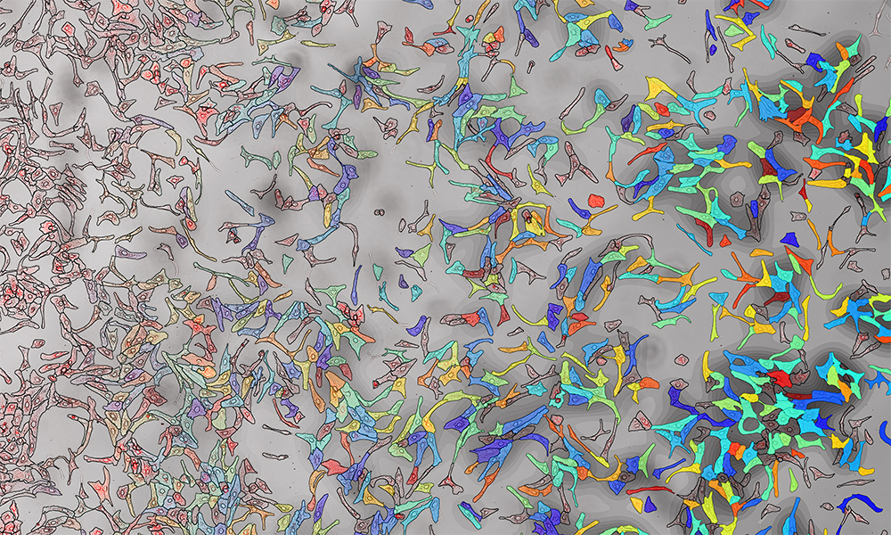
The microscope image shows liver cells grown with a surplus of oleic acid, a common fatty acid, to simulate the fatty liver condition. On the left, the intensity of redness indicates the amount of lipid accumulated by each cell, made visible with fluorescent microscopy and a chemical dye that binds to lipids. On the right, the image illustrates the amount of oleic acid within each cell (blue for low levels, red for high levels), as detected by a new method called SpaceM, developed by EMBL’s Alexandrov team. Interestingly, there are profound differences between cells. This distinct character or individuality of cells is the focus of single-cell biology, an emerging field of cellular and molecular biology.
The image was taken as part of a collaboration between the Alexandrov team and the Heikenwalder lab at the German Cancer Research Center (DKFZ).
Credit: Luca Rappez, Theodore Alexandrov/EMBL
If you have a stunning picture of your science, your lab or your site, you can submit it here.


