Chromatin cartographer
EMBL alumnus Jop Kind, this year’s John Kendrew Award winner, reflects on the imaginative questions that led him to the prize

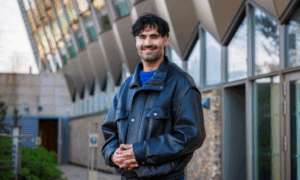
EMBL alumnus Jop Kind’s research focuses on the role of DNA organisation in genome stability, cell development and disease. Kind’s work has paved the way for the development of powerful technologies to map chromatin, or balled-up DNA, in single cells, earning him this year’s John Kendrew Award.
Chromatin was understood as a way to package and condense lots of genetic material into a tiny space. In the early 2000s, though, scientists started to understand that the way the two metres of chromatin is wrapped inside the nucleus could itself impact gene expression. Kind was gripped by this new understanding and was determined to research it more.
During his master’s internship at the Netherlands Cancer Institute, Kind came across a poster in the department of cell biology: an announcement of an EMBL conference. But he remembers most the stunning image of a castle on the poster, the Heidelberger Schloss. “I was very much into German writers and philosophers and poets from the Romantic period,” Kind remembers. “So this picture, of this castle – it really moved me.” He began searching for a lab at EMBL where he could pursue his PhD.
Hot in Heidelberg
Only a few laboratories at EMBL were working with chromatin at the time Kind saw the poster. But he joined Asifa Ahktar’s lab, which was studying the underlying mechanisms of gene expression between male and female fruit flies. Kind entered as a biochemist. At EMBL, he picked up skills in fly genetics, genomics, and bioinformatics, all in pursuit of his biggest question: How does this wrapping of DNA play a role in gene expression?
It wasn’t always easy getting to an answer. One particularly daunting challenge took place in a summer at the beginning of his PhD. It was extremely hot: up to 40 degrees on some days. Kind and his colleagues were based in temporary lab containers. “We were all in shorts and flip flops,” he says with a smile. “It definitely was not ideal.” The heat made their experiments a big mess: “We couldn’t grow bacteria because it was warmer than 37 degrees. They were in heat shock all the time!” But he kept going.
Whenever you had an idea, you could just knock on somebody’s door and talk about it
“One of my favourite parts of being at EMBL was the collaborative atmosphere,” Kind says. “Whenever you had an idea, you could just knock on somebody’s door and talk about it. We worked really intensely. But then when we went out in Heidelberg, it was also really intense!”
By the end of his PhD, Kind demonstrated one of the first links between gene expression and spatial organisation of chromatin. And it had to do not only with how the DNA was wrapped, but also its location within the nucleus. He was specifically interested in the 35 percent of DNA that was touching the interior of the nuclear membrane, or lamina. What Kind found would challenge the way scientists think about cell division and tumour growth.
Spinning yarns
For his postdoc, Kind joined Bas van Steensel’s group at the Netherlands Cancer Institute, who had developed a technique to study spatial organisation of chromatin. But what Kind wanted to do was track nuclear organisation in single cells over time, and so he fine-tuned this technique to figure out which parts of DNA touch the lamina in two methods. The first marks the DNA, the second pinpoints its identity.
Kind can explain the complexity of his lab’s work with just a hollowed-out tennis ball, red yarn, and green paint. The tennis ball, he explains, is like the nucleus. Normally the nucleus is packed with DNA, or yarn. But in order to figure out where the yarn touches the ball, Kind paints the inside of the ball. The green paint represents a sort of ink that recognises DNA and permanently stamps it. After unravelling the yarn, Kind can see exactly where it touched the “nucleus”: the contrasting green paint spots. In the lab, this “stained” DNA can be visualised on a chromosome map.
You need to be in constant reflection – especially when the big picture lies in single cells
But here is where it gets even more exciting. This molecular stamp not only sticks to mother cells, but it is also passed on to daughter cells. Kind predicted that the layout of DNA would be faithfully inherited through different generations. To his surprise, this was not the case. The DNA layout differed between individual cells of the same genetic makeup – the mother cells and daughter cells. In other words, the green paint spots were in different places among the same strands of yarn.
His work has unravelled even more questions to answer. Among them: What does this mean for the growth of specialised cells? Or tumour cells, which have a chaotic growth and gradual disappearance of nuclear structure over time? Why would one cell decide to give its genome, damaged or not, to the next generation?
Now a Junior Group Leader at the Hubrecht Institute, Kind predicts the next step will be in deciphering relationships between DNA packing and its effects on the regulation of gene activities and DNA repair. Kind remains focused. “When you are in the lab all the time, it’s easy to go into tunnel vision and veer into different directions than you were originally going,” he says. “For that, it’s important to zoom out. You need to be in constant reflection – especially when the big picture lies in single cells.”
Jop Kind will be speaking at the EMBL in Greece alumni event on the 26th of November, 2016.