
SARS-CoV-2 curtails immune response in the gut
Scientists transform human intestinal cells into ‘mini guts’ to follow infection process
EMBL scientists, together with collaborators from Heidelberg University, have provided further evidence of the gut’s role in COVID-19.
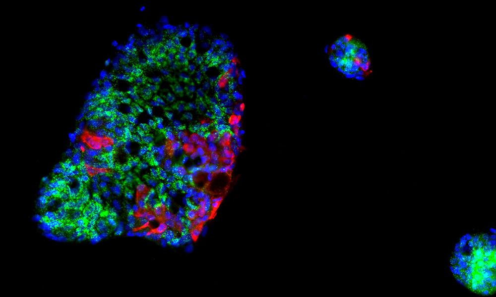
In an effort to determine the potential for COVID-19 to begin in a person’s gut, and to better understand how human cells respond to SARS-CoV-2, the scientists used human intestinal cells to create organoids – 3D tissue cultures derived from human cells, which mimic the tissue or organ from which the cells originate. Their conclusions, published in the journal Molecular Systems Biology, indicate the potential for infection to be harboured in a host’s intestines and reveal intricacies in the immune response to SARS-CoV-2.
“Previous research had shown that SARS-CoV-2 can infect the gut,” says Theodore Alexandrov, who leads one of the two EMBL groups involved. “However, it remained unclear how intestinal cells mount their immune response to the infection.”
In fact, the researchers were able to determine the cell type most severely infected by the virus, how infected cells trigger an immune response, and – most interestingly – that SARS-CoV-2 silences the immune response in infected cells. These findings may shed light on the pathogenesis of SARS-CoV-2 infection in the gut, and indicate why the gut should be considered to fully understand how COVID-19 develops and spreads.
According to Sergio Triana, lead author and a doctoral candidate in EMBL’s Alexandrov team, the researchers observed how infected cells seem to start a cascade of events that produce a signalling molecule called interferon.
“Interestingly, although most cells in our mini guts had a strong immune response triggered by interferon, SARS-CoV-2-infected cells did not react in the same way and instead presented a strong pro-inflammatory response,” Sergio says. “This suggests that SARS-CoV-2 interferes with the host signalling to disrupt an immune response at the cellular level.”
Coronaviruses, including SARS-CoV-2, cause infection by latching on to specific protein receptors found on the surface of certain cell types. Among these receptors is the protein ACE2. Interestingly, the researchers showed that the infection is not explained solely by the presence of ACE2 on the surface of the cells, highlighting our still limited knowledge about COVID-19, even after a year of tremendous research efforts worldwide.
As the disease progressed in the organoids, the researchers used single-cell RNA sequencing, which involves several techniques to amplify and detect RNA. Among these single-cell technologies, targeted Perturb-seq (TAP-seq) provided sensitive detection of SARS-CoV-2 in infected organoids. Lars Steinmetz’s research group at EMBL recently developed TAP-seq, which the researchers combined with powerful computational tools, enabling them to detect, quantify, and compare expression of thousands of genes in single cells within the organoids.
“This finding could offer insights into how SARS-CoV-2 protects itself from the immune system and offer alternative ways to treat it,” Lars says. “Further study can help us understand how the virus grows and the various ways it impacts the human immune system.”


