4 December 2024
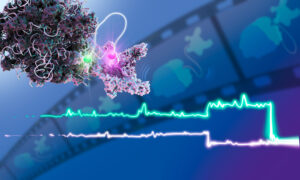
With a novel approach, EMBL scientists discovered important interactions between molecular machines, potentially offering new opportunities for drug development.
SCIENCE & TECHNOLOGY
10 September 2021
Packaged for simple installation and free use, the novel method DECODE enables researchers to reduce imaging times and increase localisation density in single-molecule localisation microscopy (SMLM).
SCIENCE & TECHNOLOGY
2021
sciencescience-technology
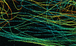
15 June 2021
As perfect as a summer night sky, these nuclear pores help calibrate a customised super-resolution microscope in EMBL’s Ries group.
SCIENCE & TECHNOLOGY
2021
picture-of-the-weekscience-technology
18 May 2021
The EMBL Picture of the Week features a series of Jurkat T cells during different stages of the activation process.
SCIENCE & TECHNOLOGY
2021
picture-of-the-weekscience-technology
15 January 2021

One of EMBL’s newest group leaders, Olivier Duss, will explore how RNA folds into functional structures and how it works with proteins to control a diverse range of activities in the cell.
LAB MATTERSPEOPLE & PERSPECTIVES
2021
lab-matterspeople-perspectives
5 January 2021
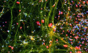
Fluorescent dyes light up a cellular community of neurons and brain immune cells (microglia), which were derived from stem cells.
SCIENCE & TECHNOLOGY
2021
picture-of-the-weekscience-technology
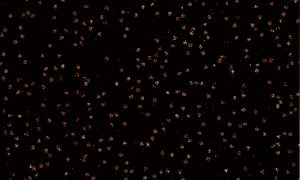
27 October 2020
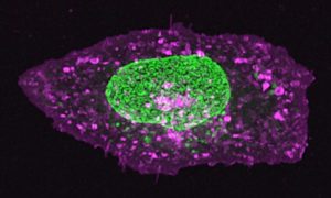
The nucleus of this cell fluoresces in bright green thanks to GFP-labelled nucleoporin proteins. EMBL scientists use engineered nucleoporins as 3D reference standards to improve super-resolution microscopy.
SCIENCE & TECHNOLOGY
2020
picture-of-the-weekscience-technology
15 September 2020
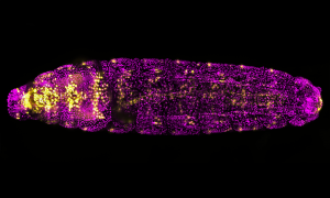
Not just another pretty fruit fly. This magenta and golden drosophila larva is lit up with a fluorescent molecule to help researchers study heart formation.
SCIENCE & TECHNOLOGY
2020
picture-of-the-weekscience-technology
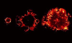
21 July 2020
EMBL scientists have created a new, realistic 3D testbed that could help achieve the goal of stopping cancers before they start by studying cancer cells as they first form.
SCIENCE & TECHNOLOGY
2020
sciencescience-technology
16 June 2020
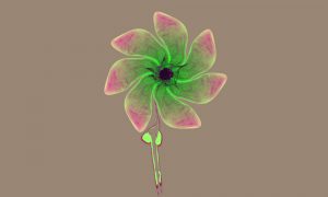
In this composite image, visual artist Mona Kakanj assembled three different biological structures in fly larvae into a flower. The original images were taken as part of a research project by Parisa Kakanj in Maria Leptin’s group.
SCIENCE & TECHNOLOGY
2020
picture-of-the-weekscience-technology
9 June 2020
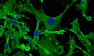
This image shows mouse embryonic fibroblasts (MEFs), their cell skeletons (green) and nuclei (blue) under a confocal microscope, photographed by Julia Hansen in the lab of Matthieu Boulard at EMBL Rome.
SCIENCE & TECHNOLOGY
2020
picture-of-the-weekscience-technology
19 February 2020
New group leader at EMBL Heidelberg employs synthetic chemistry to develop novel tools for biology
PEOPLE & PERSPECTIVES
2020
people-perspectivesscience
10 February 2020
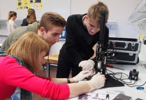
How EMBL’s ‘Microscope in Action’ introduces teenagers to the basics of fluorescence microscopy
LAB MATTERS
16 November 2010
The cells in the different parts of this video are always the same (grey), but, like actors using make-up to highlight different facial features, they have fluorescent labels that mark different cellular components in different colours: blue shows the nucleus, yellow shows tubulin (a component of…
SCIENCE & TECHNOLOGY
2010
sciencescience-technology
4 July 2010
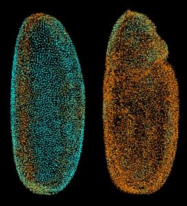
The scientists at the European Molecular Biology Laboratory (EMBL) in Heidelberg, Germany, who ‘fathered’ the Digital Embryo have now given it wings, creating the Fly Digital Embryo. In work published today in Nature Methods, they were able to capture fruit fly development on film, and were the…
SCIENCE & TECHNOLOGY
2010
sciencescience-technology
24 February 2009
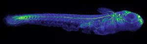
‘Useless fish with big eyes’. This is what Medaka, the name of the Japanese killifish in the pictures, means in Japan where it originally comes from. While its eyes are undeniably big, the fish has proven remarkably useful for scientists. It is a simple model organism, amenable to…
SCIENCE & TECHNOLOGY
2009
sciencescience-technology
No results found