Digital Embryo gains wings
Now possible to film development of fruit fly and of zebrafish’s eyes and brain
The scientists at the European Molecular Biology Laboratory (EMBL) in Heidelberg, Germany, who ‘fathered’ the Digital Embryo have now given it wings, creating the Fly Digital Embryo. In work published today in Nature Methods, they were able to capture fruit fly development on film, and were the first to clearly record how a zebrafish’s eyes and midbrain are formed. The improved technique will also help to shed light on processes and organisms, which have so far been under-studied because they could not be followed under a microscope.
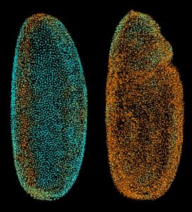
“Non-transparent samples like the fruit fly embryo scatter light, so the microscope picks up a mixture of in-focus and out-of-focus signal– good and bad information, if you like,” says Ernst Stelzer, whose group carried out the project at EMBL. “Our new technique enables us to discriminate between that good and bad information, so it allows us to record organisms which have so far been poorly studied, because of their unfortunate optical properties.”
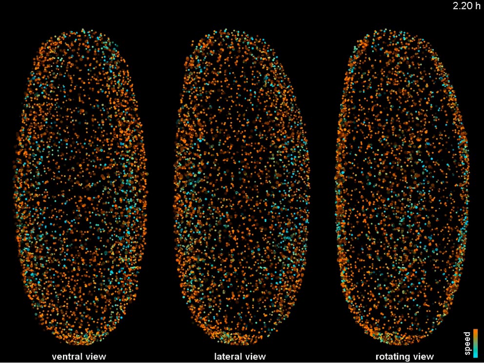
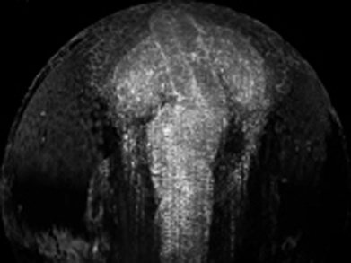
Philipp Keller, who co-led and conducted the work, and Ernst Stelzer overcame the difficulties caused by thick, opaque samples, by shining patterns of light on them, instead of the usual continuous light sheet. This generates an image with alternating light and dark stripes, unless the light bounces off the sample and changes direction, in which case this stripy pattern will be blurred. By taking multiple images of different phases of the light pattern, and combining them, a computer can filter out the effects of scattered light and generate an accurate image of the sample, thus enabling scientists to record images that were previously unobtainable.By combining this approach with imaging along different angles, the scientists were able to obtain three-dimensional movies of the developing fruit fly embryo in spite of the fact that it is almost opaque.
The EMBL scientists were also able to extend their recordings of zebrafish development to an unprecedented level. They took around one million images to capture the first three days of zebrafish development from three different angles, generating films in which the formation of the animal’s eyes and midbrain are clearly visible.
“Of course, getting such good images is nice for the human observer, but it’s particularly crucial for computational analyses, like tracking cell movements and divisions as we do in the Digital Embryo,” says Philipp Keller, now at the Janelia Farm Research Campus of the Howard Hughes Medical Institute in Ashburn, VA, USA.
The work was done in collaboration with scientists at the University of Heidelberg, Germany and the Sloan-Kettering Institute in New York, USA.
All data, images and videos are freely available online, alongside the data from the digital embryo, at www.digital-embryo.org.
For more information about the first Digital Embryo, click here.


