
Glow-in-the-dark cell skeletons
Skeletons provide shape, structure, and stability. For these reasons, cells also contain a skeleton – one that’s made of different proteins. In contrast with the bony skeletons of whole organisms, cell skeletons (also called cytoskeletons) are highly dynamic. The proteins within them can be flexibly rearranged, which helps cells to change their shapes. The cytoskeleton also enables cells to transport cargo, such as other proteins, in a coordinated manner through the cell, with the cellular skeleton acting as a sort of conveyor belt.
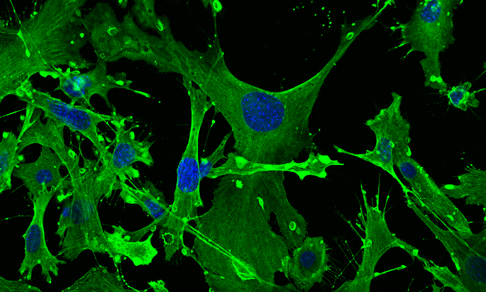
This image shows mouse embryonic fibroblasts (MEFs) and their cell skeletons under a confocal microscope. The cells were stained with fluorescent antibodies against the actin protein, which is part of the cell skeleton (shown in green). The cell nuclei were made visible with a fluorescent dye called DAPI, which binds to DNA (shown in blue). Julia Hansen, a Biochemistry master’s student at Heidelberg University, took this image while testing various antibodies with MEFs during her internship in the Boulard group at EMBL Rome. While light is required to excite electrons and induce fluorescence, the latter is usually much lower in intensity than the excitation light. Hence, emission filters in the microscope are used to block the excitation light, enabling us to see the fluorescence on a dark background.
Credit: Julia Hansen/EMBL


