
Fruit fly with a heart of gold
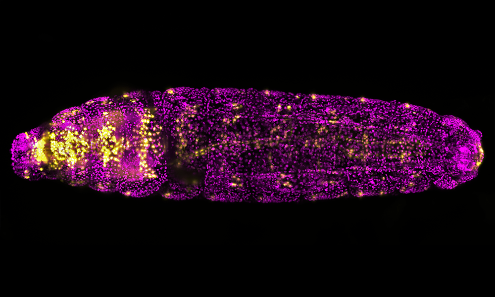
Pretty in pink, this fruit fly larva is much more than just another pretty fly. Its heart seems almost like gold, thanks to a fluorescent molecule – or fluorophore – that marks cells involved in heart formation.
During the fruit fly’s embryonic development, its tubular heart is formed on its back. Researchers in the group of Eileen Furlong at EMBL are interested in the origin of the first heart-forming cells. They created a fly line that expresses a fluorophore able to mark those specific cells. Together with researchers in Lars Hufnagel’s group, they can record the early development of the fruit fly embryo and trace back the steps of heart formation with the help of a sophisticated light-sheet microscope (MuVi-SPIM). This is a specialised type of microscope that was developed at EMBL.
This image shows an early Drosophila larva with the nuclei of heart-associated cells highlighted in yellow, and other nuclei shown in magenta.
Credit: Dimitri Kromm, Enrico Cannavò/EMBL
If you have a stunning picture of your science, your lab or your site, you can submit it here.


