Cellular fireball or immune cells?
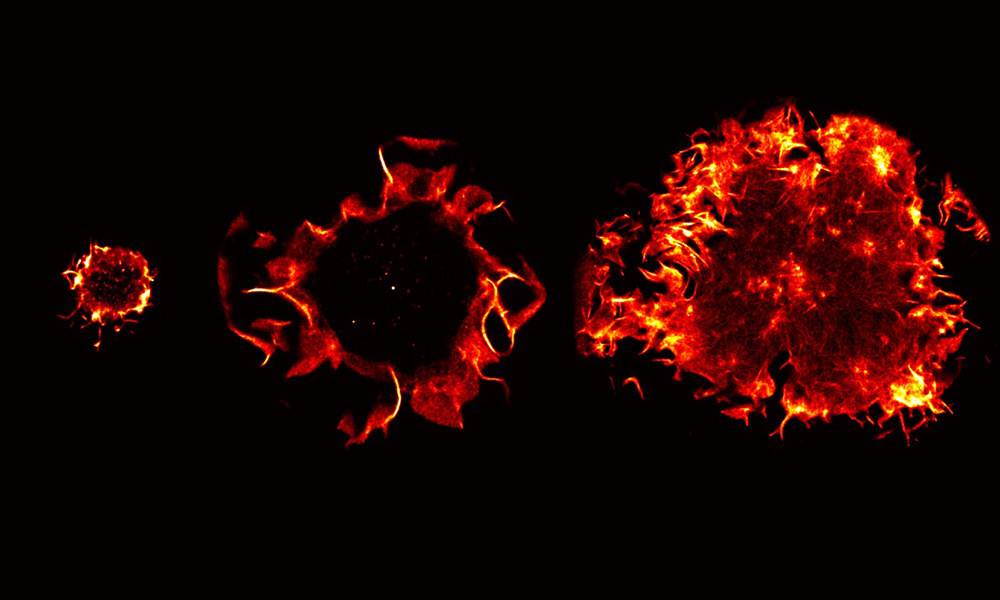
These images of T cells may look a bit like encroaching fireballs, but that’s due to the dazzling fluorescence used to study them. Our EMBL Picture of the Week features a special type of T cell called a Jurkat cell.
T cells are white blood cells that are part of the immune system, designed to keep us healthy. Jurkat T cells, an immortalised line of human T cells, have become particularly useful for studying a type of leukaemia called T-cell acute lymphoblastic leukaemia, a condition where immature white blood cells crowd out useful, mature ones. Immortalised cell lines are created from cells that would not normally proliferate indefinitely, but due to mutation they are now able to do so, continuing to grow and divide for prolonged periods in a laboratory setting.
The technology that makes these cells visible with such clarity is single-molecule localisation microscopy, a super-resolution technique that helps researchers like Angelica Maria Estrada Pacheco in EMBL’s Ries group to explore how these immune cells change their structure upon activation, meaning when they encounter an intruder or antigen and prepare to respond to protect the body.
This photo series features three Jurkat T cells during different stages of the activation process. From left to right, we move from non-activation, suspected activation in progress, and finally a cell after being activated, having increased in size.
Single-molecule imaging techniques include fluorescence microscopy approaches that use fluorescent tags to detect and analyse individual molecules. In this case, researchers use the approach to unravel how the network of a protein called actin in T cells plays a role in helping the cells in their search for antigens. The actin network also facilitates the downstream signalling cascade that allows the activation of T cells into effector T cells. The cells become considerably larger upon activation, and this seems mostly due to a reorganisation of the actin network.
Credit: Angelica Maria Estrada Pacheco/EMBL
If you have a stunning picture of your science, your lab or your site, you can submit it here.