
It’s like a party in your brain
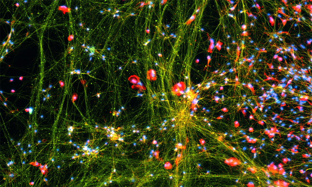
Looking a lot like flashy lights and streamers left over from New Year’s Eve festivities, these decorations are hardly that. This image represents work in EMBL’s Zaugg and Noh Groups in which postdoc Mikael Marttinen used human stem cells to generate neurons and brain immune cells (microglia) that were co-cultured – growing them together in a dish. Not only is this a pretty picture, it represents an advance that will help EMBL researchers study neurodegenerative diseases.
The green stain (Alexa Fluor 488) shows a protein known as class III beta-tubulin, which forms long (and apparently flashy) filaments known as microtubules, and is an explicit marker for neurons. Those blingy pink or red dazzlers (getting their colour from a dye called Alexa Fluor 594) are another kind of protein known as TMEM119, which lights up microglia – the brain’s immune cells. Lastly, a fluorescent blue stain called DAPI lights up cell nuclei. This image shows that Marttinen was able to generate a co-culture system with both neurons and microglia, making it a good model system to study diseases like amyotrophic lateral sclerosis (ALS), Parkinson’s disease, or Alzheimer’s at the molecular level.
Credit: Mikael Marttinen/EMBL
If you have a stunning picture of your science, your lab or your site, you can submit it here.


