
From fly to flower
Parisa Kakanj, a postdoc in Maria Leptin’s group at EMBL Heidelberg and the University of Cologne, recently developed and published a technique to study wound healing in living fruit fly larvae. The larvae already have highly developed organs, yet their body is still transparent, which allows scientists to observe organs inside the living animal under the microscope.
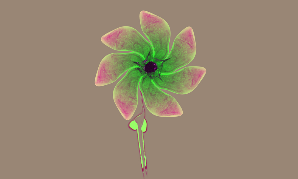
When visual artist Mona Kakanj saw the images her sister created as part of her study, she realised that they could be repurposed to create a work of art. In this composite image, she assembled three different biological structures in fly larvae into a flower.
The flower’s petals are made up of seven rotated images of part of a wing imaginal disc. The imaginal disc is a structure of epithelial cells, which will later turn into the fly’s wings when the pupa undergoes metamorphosis. The petal’s centre is made up of an image of a healing wound. The stem is composed of the organ that the larvae use to sense their surroundings and adapt their movement accordingly. Parisa expressed green fluorescent proteins in the larvae to visualise them under the microscope. The different colours were added afterwards. The original images were published as part of Parisa’s recent paper in the journal Nature Protocols.
Credit: Parisa Kakanj/EMBL, Mona Kakanj


