Picture Release
Images of Medaka juveniles taken with a Digital Scanned Laser Light Sheet Fluorescence Microscope.
‘Useless fish with big eyes’. This is what Medaka, the name of the Japanese killifish in the pictures, means in Japan where it originally comes from. While its eyes are undeniably big, the fish has proven remarkably useful for scientists. It is a simple model organism, amenable to genetic techniques, easily grown in the lab, but at the same time it shares many molecular processes with higher vertebrates. Particularly its embryos and juveniles are widely used to address questions about embryonic development.
Click to download High resolution .tiff
Click to download High resolution .tiff
Click to download High resolution .tiff
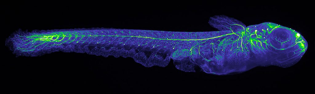
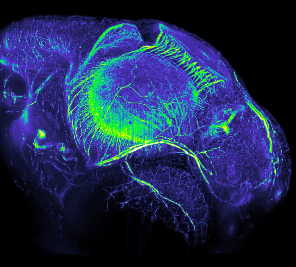
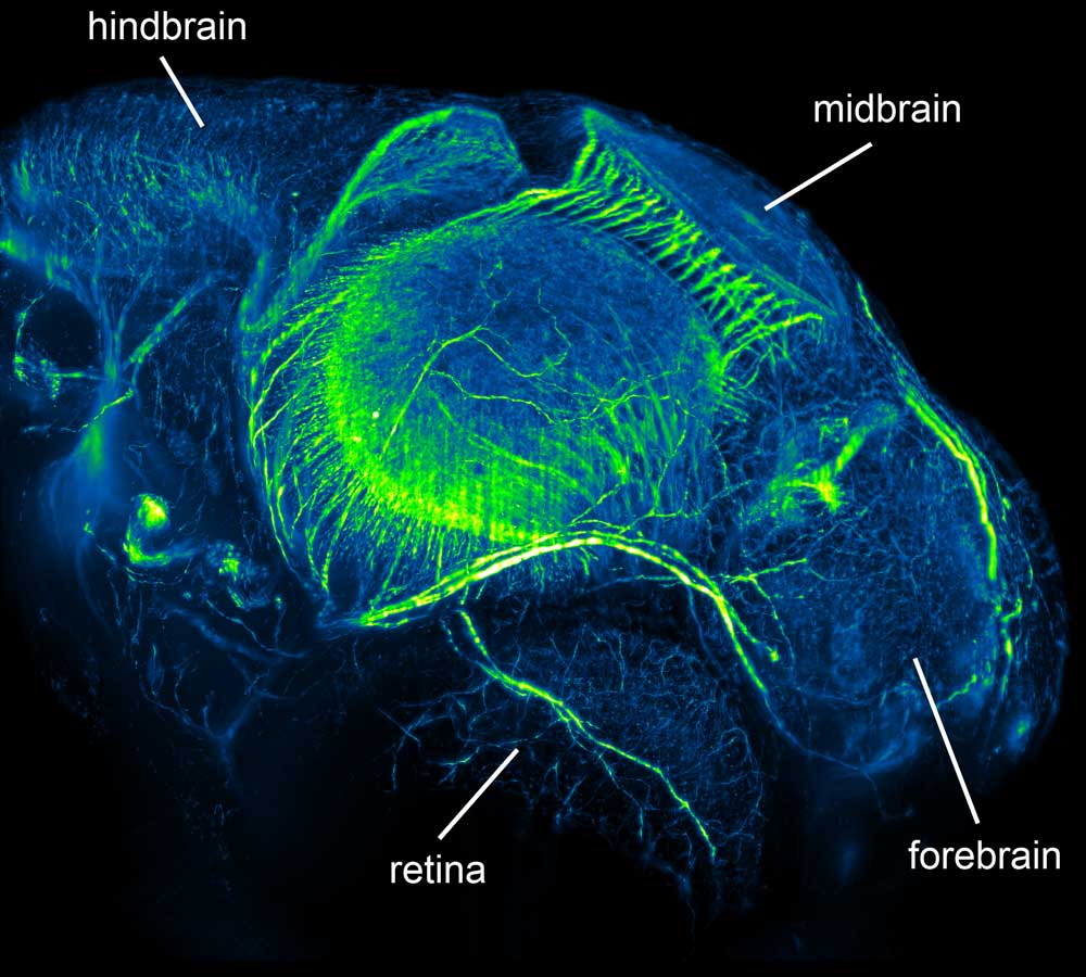
The two images of Medaka juveniles, a full body shot and a head close- up, were taken by Philipp Keller, from the lab of Ernst Stelzer at the European Molecular Biology Laboratory (EMBL), with a newly developed microscope called Digital Scanned Laser Light Sheet Fluorescence Microscope. The specimens were prepared by Lazaro Centanin and Annette Schmidt from Jochen Wittbrodt’s lab at EMBL. The first image shows a Medaka juvenile at the age of 10 days. It is 5 mm long (2.5 times magnified) and the staining shows its developing brain, eye and spinal cord. Zooming in 20 times on the head (700 µm x 600 µm) of a slightly younger Medaka reveals (from right to left) forebrain, retina of the right eye, midbrain and hindbrain at the age of 4 days.


