Bio-SAXS is very versatile – it can be used for samples in almost any form, e.g. liquid or solid.
Swordfish sword under X-rays: SAXS explained
Learn how scientists use X-rays to study bones at a molecular level
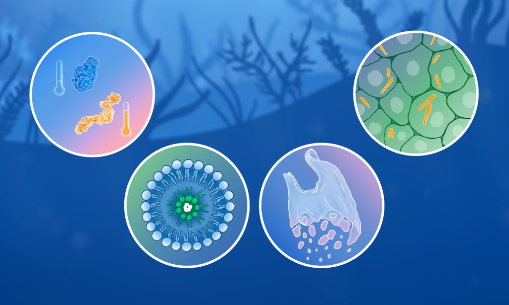
Animated infographic. Scientists use X-ray techniques, such as bio-SAXS, to study molecular structures in various settings, even in the bone that forms the swordfish’s sword. Credit: Dorota Badowska, Cy Jeffries, Joana Carvalho, Isabel Romero Calvo, Da In Wi, Tabea Rauscher /EMBL
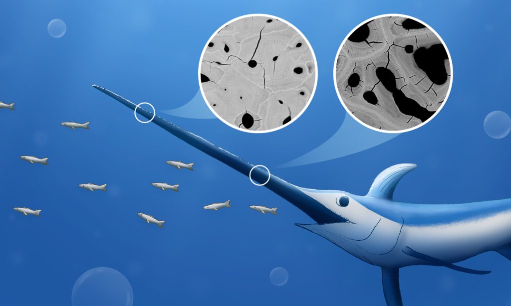
Did you know that swordfish use their long sword as a tool for hunting? Rather than using it like a sword, they use it as a club to stun their prey. However, despite being under heavy load during hunting, it doesn’t break easily.
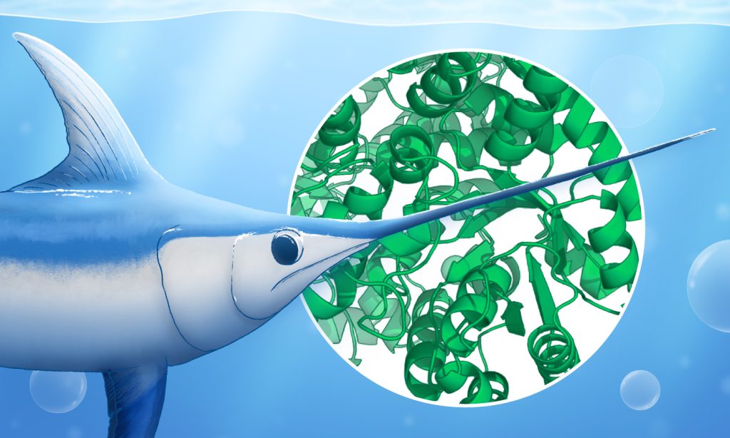
Scientists explore this material strength using various experimental techniques, such as SAXS. Because the swordfish sword is in many ways similar to the bones of older human adults, it can help in understanding human bone ageing.

Bio-SAXS: biological small-angle X-ray scattering
Bio-SAXS lets scientists determine the shape and dynamics of proteins and other bio-molecules using X-rays.
X-ray experiments take place at specialised testing stations called beamlines in synchrotrons.
During a bio-SAXS experiment, X-rays are shot at a biological sample and get scattered in a unique pattern as they pass through it. This pattern depends on the types of atoms and how they are arranged inside the sample.
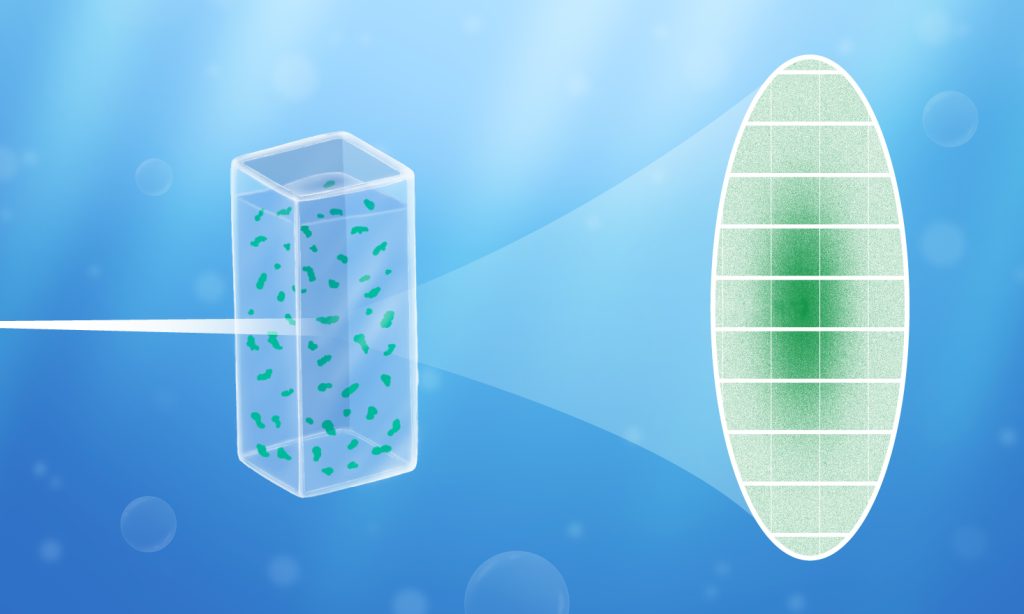
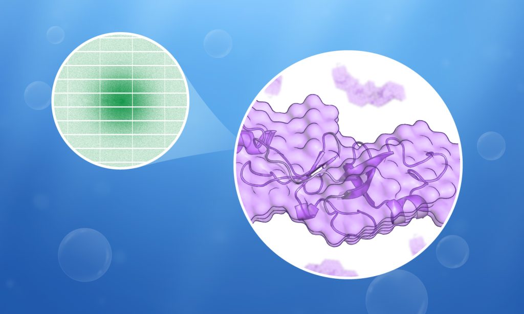
Scientists then analyse the scattering pattern to ‘peek inside’ a material to work out the 3D structures of the molecules.
Bio-SAXS is very versatile – it can be used for samples in almost any form, e.g. liquid or solid. It allows studying how molecules change their structure in different conditions, and how they interact with each other. The molecular structures and interactions are what give materials their unique properties.
Understanding human bone ageing using the swordfish sword
Bio-SAXS has previously enabled scientists to examine the structure of mineral particles in the swordfish sword at both the young bone at its base and the old bone at its tip. Studying this could help us understand better why and how bones in older adults deteriorate with time.
Other applications of bio-SAXS
Bio-SAXS helps scientists address other questions as well:
- Detect crystals growing inside cells
Download the full-size SAXS infographic
The infographic is available under the CC-BY-SA 3.0 IGO license.
This infographic was produced thanks to the collaborative effort of several people:
Concept and text: Dorota Badowska and Cy Jeffries/EMBL
Visual concept and design: Joana Carvalho, Isabel Romero Calvo/EMBL
Animation: Da In Wi/EMBL
Creative lead: Tabea Rauscher/EMBL
Scientific consultations: Clément Blanchet and Cy Jeffries/EMBL, Imke Fiedler and Felix Schmidt/UKE
Source article(s)
Versatile sample environments and automation for biological solution X-ray scattering experiments at the P12 beamline (PETRA III, DESY).
Journal of Applied Crystallography 12 March 2015
10.1107/S160057671500254X
On the Origins of Fracture Toughness in Advanced Teleosts: How the Swordfish Sword's Bone Structure and Composition Allow for Slashing under Water to Kill or Stun Prey
Advanced Science 2 May 2019
10.1002/advs.201900287


