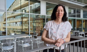
Welcome: Wojciech Galej
New group leader at EMBL’s Grenoble site investigates RNA-protein complexes involved in gene expression

Wojciech Galej is very new to EMBL, but his lab overlooking the Chartreuse Mountains is filling up quickly. Soon his group will use state-of-the-art methods in structural biology and biochemistry to investigate the structure of large RNA-protein complexes.
How did the move to EMBL go?
So far, so good. It’s been an exciting time! In my first week here I flew to Shanghai to give a presentation, so the pace has been quite frenetic. Things in the lab are moving quickly – we have already started doing first experiments together with my newly recruited research technician, Estelle Marchal.
What is your group working on?
The structure of biological molecules plays a major role in determining their function. My group uses structural biology methods to study the molecular bricks that, when assembled, form the basis of one of the most complex yet fundamental cellular machines: the spliceosome.
The first step of gene expression is a process called transcription. Eukaryotic genes are transcribed into pre-messenger RNA. This pre-mRNA is punctuated by regions that code for proteins – exons – and regions that do not code for proteins – introns. The spliceosome acts like a molecular editor: it cuts out the introns and pieces the exons back together. This process creates a molecular blueprint for making a protein.
What sorts of tools do you use?
The spliceosome is extremely complex and dynamic. By using a combination of wet lab experiments and cryo-electron microscopy (cryo-EM), our goal is to understand how its different parts assemble and work together.
A lot of the time I’m at the bench doing wet lab experiments – cloning genes, purifying proteins, and making some biochemical assays. Then we move on to the microscope and image our samples. This process is followed by extensive computational data processing and model building.
When I was a PhD student, I began studying the structure of the spliceosome using X-ray crystallography. My project was a success – I had solved a 3D structure of a protein that was positioned at the very centre of the whole complex. However, the machinery of the spliceosome is very dynamic, and X-ray crystallography limited us to using samples that weren’t too big or too flexible. In my postdoc we switched to using cryo-EM, which allows us to look at structures that are much more dynamic and flexible. The setup we have on the European Photon and Neutron Science Campus in Grenoble, along with an easy access to both the ESRF synchrotron and a soon-to-arrive cryo-electron microscope, allows us to do cutting edge research and stay at the forefront of structural biology.
What skills are you looking for when recruiting?
There are particular technical skills I am looking for. I’m looking for scientists that are strong in biology and chemistry, but at some point I would also like to recruit someone with experience in computer science and physics. Combining the knowledge gained from these areas of expertise will give us a 360° view on the problems we investigate. The most important thing is to be passionate about science; everything else can be learnt while working on a particular project.
Can you recall a particularly cool experiment that you’ve done?
In every structural biology experiment there is that moment of seeing your structure for the first time. It is an amazing feeling to be the only person in the world to know something that others don’t. I have to confess that sometimes I like to prolong that moment by not telling my colleagues straight away!
It is an amazing feeling to be the only person in the world to know something that others don’t
Solving the structure of Prp8 was a very memorable experience for me – partly because it was a project that was around in the lab for more than a decade, and partly because it was regarded by many as almost impossible. Thanks to fantastic support from my supervisor Kiyoshi Nagai and senior lab scientist Andy Newman, not only did we crystallise it, but we also determined the structure at atomic resolution, providing an incredibly detailed 3D view of this protein. Unexpectedly we also found an evolutionary link to group of proteins that offer insight into how such a complex structure could have evolved.
The process of solving a molecular structure is much like solving a jigsaw puzzle. The only difference is that while we know what the pieces look like, we don’t really know the final image we are aiming to put together – and the pieces often don’t fit together perfectly. So sometimes we need to extrapolate and be creative to understand what the protein looks like and investigate its function.
What got you into science?
I was quite a keen experimentalist as a youngster, and I had a little chemistry lab at home. Initially this was more of a hobby than something I saw turning into a career. But while I was initially intending to pursue a career in medicine – like my father and my brother – these early experiments and some inspiring teachers I met along the way had a profound impact on me. I switched my focus to experimental science. It’s been a challenging and very rewarding career path, and I have never looked back!


