EasyGrid: towards better automation in cryo-electron microscopy
EMBL Grenoble researchers have developed unique instruments automating sample preparation and quality control for cryo-electron microscopy
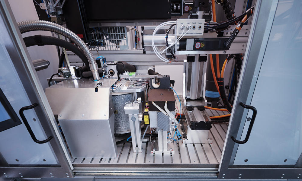
The EasyGrid instrument installed at the EMBL Imaging Centre. Credit: Joseph Franciosa/EMBL
Summary
While cryo-electron microscopy is a powerful method for determining the three-dimensional structure of proteins, preparing high-quality samples for it remains challenging.
The Papp Team at EMBL Grenoble has developed two prototypes to automate sample preparation and quality control: EasyGrid and EasyGrid Control, as described in a recent Nature Methods publication.
These unique instruments can be used across scales for cryo-electron microscopy and tomography, as well as X-ray nano-imaging.
EasyGrid Control will soon be offered as a service to the scientific community and prepared to be commercialised.
Cryo-electron microscopy (cryo-EM) can help scientists determine the three-dimensional structure of proteins in unprecedented detail. Jacques Dubochet, former Group Leader at EMBL, shared the 2017 Nobel Prize in Chemistry with Joachim Frank and Richard Henderson for the development of this technique, which led to the ‘resolution revolution’ in structural biology.
Despite recent advances in cryo-EM methods, preparing high-quality samples remains challenging. The Papp Team at EMBL Grenoble, which specialises in creating innovative instrumentation for structural biology, is currently addressing this problem.
The team developed two unique systems, EasyGrid and EasyGrid Control, which they describe in a new Nature Methods publication. These platforms automate the preparation of high-quality samples for studies across scales – from single particles to whole cells – and can be adapted to different techniques: cryo-EM, cryo-electron tomography (cryo-ET), and X-ray nano-imaging.
What is cryo-electron microscopy?
Adopted by structural biologists as a complementary technology to nuclear magnetic resonance (NMR) and X-ray crystallography, cryo-electron microscopy allows scientists to study the structure and function of macromolecular assemblies (proteins and protein complexes) at the near-atomic level.
The method involves passing a beam of electrons through a vitrified sample (a sample that has been rapidly frozen at very low temperatures). Researchers then obtain two-dimensional projections of macromolecules that can be reconstructed as 3D models using computational algorithms.
Improving replicability and sample quality
The primary limitation to preparing high-quality vitrified samples is the manual preparation of samples, which requires careful handling and suffers from a lack of reproducibility in ice thickness and ice quality. If the ice layer is insufficiently thin or full of ice crystals, the electron beam cannot penetrate it or deviates due to diffraction, rendering the sample unsuitable for further imaging.
In addition, there is no way to easily pre-screen and check the quality of vitrified samples before viewing them using a cryo-electron microscope. These limitations make for poorly reproducible, lengthy, and often costly experiments.
The EMBL Grenoble Instrumentation team, which has previously automated processes in X-ray crystallography – now used in most synchrotrons worldwide – took on the new challenge of improving cryo-EM sample preparation processes in 2017.
After several years of iterations on a first prototype, the team, led by Gergely Papp, created EasyGrid – a fully automated and modular platform for high-throughput sample vitrification. In parallel, they developed EasyGrid Control – a unique instrument capable of automatically screening samples prepared with either EasyGrid or traditional methods to check their quality before use in advanced microscopy.
“With EasyGrid and EasyGrid Control, the whole process line, from the loading of the grid to the screening, is done autonomously,” explained Vic Armijo, mechatronics engineer in the Papp Team working on this project since 2018, and co-first author of the publication. “This leads to a more repeatable process and allows us to optimise samples faster and measure their quality, so we only load the best grids into the electron microscope.”
Automated sample preparation across scales
Another important aspect of EasyGrid is the use of different sample dispensing and vitrification methods from those currently offered by other sample preparation techniques.
The system uses pressure waves to spread the sample across the grid’s surface, enabling reliable liquid-layer thinning for single-particle analysis. It can also do this for samples comprising cells cultured directly on the surface of the sample holder grid or in suspension, dispensed with automated pipettes.
Additionally, while conventional preparation methods usually involve immersing the grid in liquid ethane, the Papp Team has developed an ethane jet-based vitrification system using bare grids (grids that are not mounted in an autogrid cartridge, a kind of circular copper frame). “This dramatically improves the cooling rate, enabling vitrification deeper within the cell, including the nucleus,” said Armijo. Hence, this sample preparation method is also advantageous for in situ (in the context of the cell) studies using other imaging technologies, such as cryo-electron tomography and X-ray nano-imaging.
EasyGrid was developed thanks to a close collaboration with researchers from EMBL sites with expertise in structural and cell biology (Grenoble, Hamburg, and Heidelberg) and partner institutes in France, such as Inserm, Institut Pasteur, Sorbonne Université, and the Institute of Advanced Biosciences (IAB), who tested, provided feedback, and validated the prototypes.
Throughout the project, the Papp Team obtained high-resolution maps of several macromolecular complexes, including apoferritin, yeast ribosomes, guanidinase, the INO80–nucleosome complex, and pentamers of the bacterial rhodopsin KR2. The team also demonstrated a sevenfold improvement in the vitrification quality achieved within the nuclei of large human cells, compared with traditional plunge-freezing methods.
“Such a platform is an important game-changer not only in the quest for reliable automated cryo-preparation for high-throughput analysis by cryo-EM, but also for other types of atomic-resolution imaging of biological samples preserved in their native hydrated environment,” said Sylvain Bohic, senior researcher at Inserm, France.
“We evaluated the platform within our project on intracellular metal imaging, focusing both on the fate of theranostic nanoparticles [small particles that can be used simultaneously for medical diagnostics and therapy] and on how metal ions influence protein aggregation,” added Bohic. “The method delivered robust, reproducible cryo preparation, embedding cells in a very thin, homogeneous layer of amorphous ice. It also adapts seamlessly to our custom sample supports – square silicon nitride windows used for synchrotron cryo X-ray fluorescence nano imaging – resulting in superior image quality.”
Technology transfer and service to the scientific community
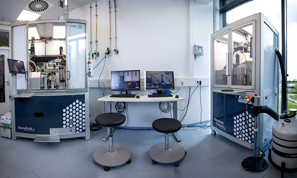
The EasyGrid and EasyGrid Control prototypes developed at EMBL Grenoble have been transferred to the EMBL Imaging Centre (EMBL IC) in Heidelberg.
“EasyGrid and EasyGrid Control represent a significant advancement in cryo-EM technology, which will dramatically accelerate the optimisation process for both single-particle analysis and cellular cryo-electron tomography,” said Simone Mattei, Electron Microscopy Team Lead at EMBL IC. “The integration of these versatile platforms at the EMBL IC will provide researchers with unprecedented control over sample vitrification parameters and enable high-throughput screening of specimens before valuable microscope time is allocated, ultimately enhancing the efficiency and success rate of structural biology projects.”
EasyGrid (left) and EasyGrid Control (right) instruments at the EMBL Imaging Centre. Credit: Stuart Ingham/EMBL
The Papp Team is now working on merging these two technologies. “Our instruments are now mature enough to be recombined in one single machine, so we are currently putting together a third version of EasyGrid at EMBL Grenoble, which will combine sample preparation and quality control,” explained Papp.
The three machines at EMBL Heidelberg and EMBL Grenoble will be available as a service to EMBL researchers and to the scientific community through Instruct-ERIC.
The team is also setting up a new EMBL Grenoble spin-off company, which will be led by Armijo, to commercialise their prototypes with the support of EMBLEM, EMBL’s technology transfer partner and commercial arm. “The first instrument available on the market will be a version of EasyGrid Control with improved resolution and an optional fluorescence microscopy module,” explained Papp. “Later, a simplified EasyGrid dedicated to cell vitrification should follow the same path. We are already thinking about how to simplify this sample preparation system to make it commercially accessible.”
Funding and support
The development of the EasyGrid instruments has benefitted from several funding sources:
EU project: Fragment-Screen (grant agreement ID: 101094131)
EU project: IMAGINE (grant agreement ID: 101094250)
ARISE programme (Marie Sklodowska-Curie, grant agreement number 945405)
iNEXT-Discovery (project number 871037)
Joint support via Marie Skłodowska-Curie Actions (101028297), Biomedicum Helsinki Foundation, the Orion Foundation and The Finnish Cultural Foundation
EMBL Interdisciplinary Postdoc Programme (EIPOD) under Marie Curie Actions COFUND.