Online Magazine of the European Molecular Biology
Laboratory
Visualising biology: new tools of the trade
EMBL researchers are pushing the frontiers of big data analysis in biological imaging, allowing scientists to gain a many-layered and multidimensional view of organisms, tissues, and cells in action.
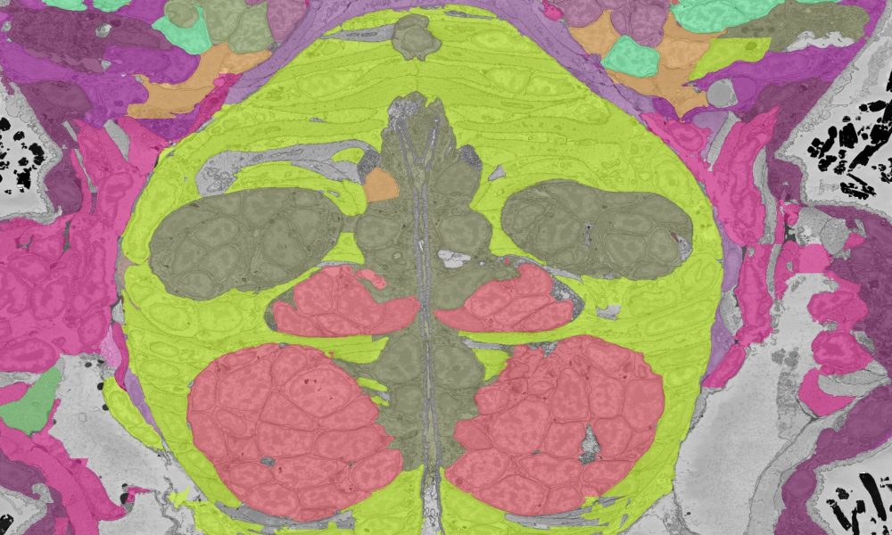
A section of electron microscopy volume of a Platynereis larvae. Credit: Arendt group
Biological imaging reveals to us the wonderful inner worlds of living organisms, bringing into sharp focus all their quirks, oddities, and moving pieces. EMBL has long been a world leader in this field, spearheading advances in imaging technology at the same time as making imaging services accessible to the wider scientific community.
With progress in imaging technology, however, comes the problem of handling the huge datasets that such methods inevitably produce. Researchers across EMBL have been collaborating to find a solution to this 21st-century problem, and the tools they are developing will help researchers across the world share, analyse, and collaborate on imaging data for years to come.
The problem of big data in microscopy
In addition to letting us peek inside organisms, bioimaging helps us understand how they function. They also let us track the way these functions change in response to disease states or environmental challenges. From the 16th-century compound microscopes created by Dutch spectacle-makers to today’s state-of-the-art cryo-electron microscopy facilities, bioimaging technologies share a common purpose: to allow us to see deeper into the fundamental mechanisms of living systems.
The last few decades have seen explosive growth in the capabilities of such technology systems. In recent years, EMBL researchers have pioneered techniques that, among other applications, let us decrypt molecular structures inside cells, combine imaging with next-generation sequencing methods, and measure the mechanical properties of developing embryos. In addition to increasing the resolution of optical microscopy beyond what was once thought possible, scientists worldwide have made significant advances in combining different modalities of bioimaging in the form of correlative microscopy.
Correlative microscopy allows researchers to place layers of information on top of each other. While one technique, e.g. electron microscopy, might show us cellular ultrastructures, another, like fluorescence microscopy, might help us pinpoint the location of various proteins. By combining such information, researchers can gain significant insight into biological functions.
However, the ever-increasing resolution and scope of imaging technologies result in ever-expanding datasets, with file sizes ranging into the order of terabytes. This, unfortunately, makes it impossible to open and view such files on an ordinary computer, requiring the use of extensive computing resources. Additionally, extracting biological meaning out of such colossal datasets can be a time-consuming as well as error-prone endeavour.
Mobilising MoBIE
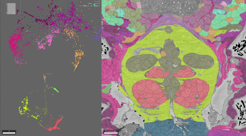
Scientists can explore cell types and tissues using the MoBIE interface. The image above shows a scatter plot depicting single cells based on their morphological properties and a section of electron microscopy volume of a Platynereis larvae, with different colours representing automatically determined animal tissues. Credit: Valentyna Zinchenko/EMBL
In order to deal with this challenge, EMBL scientists Christian Tischer, Yannick Schwab, Anna Kreshuk, and Detlev Arendt, began a collaboration in 2018 to build a tool that would allow researchers across the world to share and view such multifaceted datasets on simple computing systems.
The result of their efforts was MoBIE – a software tool that allows scientists to handle large imaging datasets, as well as share and analyse them collaboratively. It can help scientists visualise data in multiple dimensions (e.g. in 2D, 3D, and 4D) and integrate data from many different domains of biology.
“MoBIE enables the exploration and sharing of really big correlative image datasets,” said Tischer. “It builds on existing technologies, such as BigDataViewer and next-generation image file formats, and adds features for combining large heterogeneous images and corresponding segmentations into easily browseable projects.”
First described in a publication in Nature Methods earlier this year, MoBIE allows users to seamlessly stream data from a remote server, and share “views” of imaging datasets with each other. It is also free to download for researchers worldwide. In addition to electron microscopy data, MoBIE can be used to integrate data from fields ranging from gene expression to X-ray imaging.
According to Schwab, this development is also significant for imaging services. “A tool like MoBIE can enable smooth communication with users. By using MoBIE, data producers and users can interact with the datasets after and even during the production process,” he said.
Machine learning to decode cellular signatures
One of the early adopters of this tool was Detlev Arendt, whose group studies the evolution of the nervous system by using the worm Platynereis as a model system. In 2021, the researchers created the first multimodal cellular atlas combining electron microscopy and expression data for an entire animal, which was made available to the global research community via the MoBIE technology.
The creation of this atlas was enabled by volume electron microscopy, which is a method wherein electron microscopy techniques are applied to ‘large’ volumes to generate a three dimensional view of a cell, tissue – or an entire organism, in the case of Platynereis.
“This approach exemplified this important transition to go from serial sections into visualising the entire volume of an animal”, said Arendt. “You can almost think of it now as a virtual reality space – using your cursor, you can travel through this volume, and find things that have never been seen before.”
However, with such a huge dataspace full of so many unknowns, correctly annotating cells or tissues can be an important challenge – one that can take hours of painstaking manual labour.
To solve this problem, in another new study, published in eLife, Arendt and Kreshuk describe a novel approach to analysing this dataset and extracting meaningful biological information from it. By using a neural network-based deep learning approach, the team automated the process of identification of cells, cell types, and tissues at organism-scale by identifying distinct morphological features. The method, aptly named ‘MorphoFeatures’, could group similar cells and cell types – classifications that could be verified by using gene expression data.
During the whole life-cycle of the MorphoFeatures development, MoBIE was used to explore the data and visually validate the method, as well as discover and study morphological points of interest. Now, researchers in the world can now take a look at the data and apply the MorphoFeatures method themselves.
“This is a pioneering study for this kind of analysis in volume electron microscopy, and we want to generate many more such analyses,” said Arendt. “This is a new field that is just starting and visualisation tools such as MoBIE are critical to do the same for many more organisms in many different ways.”
Volume electron microscopy at EMBL
Volume EM (vEM) was first introduced at EMBL around 10 years ago by the Electron Microscopy Core Facility and the Schwab team. In recent years, this versatile technique has seen widespread use in many life science fields. In 2022, EMBL researchers contributed to a primer on vEM published in Nature Review Methods. They are also helping organise the first Gordon Research Conference on vEM, to be held in July 2023. In addition to pioneering the use of vEM for answering fundamental biological questions, EMBL has also been a leader in integrating vEM with other imaging technologies, especially 3D fluorescence microscopy and X-ray imaging.
Looking ahead
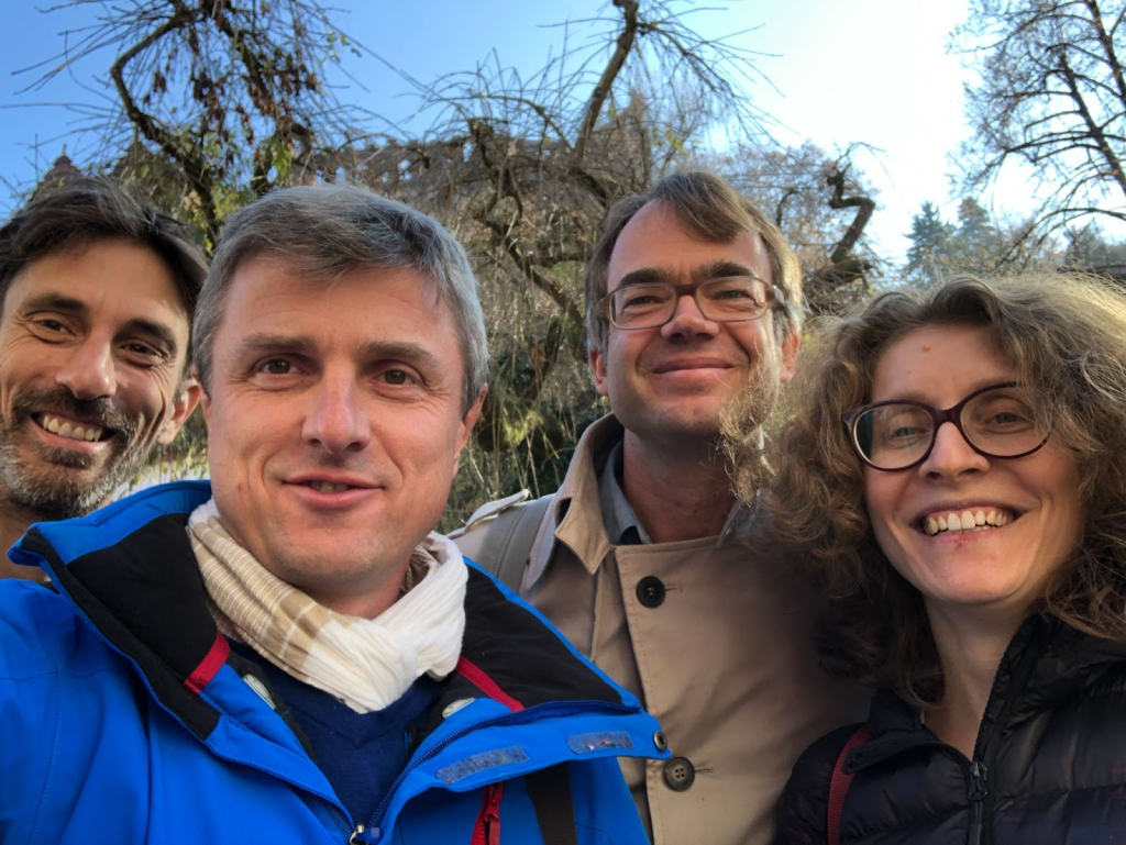
Caption: The four collaborators in 2018. From left to right: Christian Tischer, Yannick Schwab, Detlev Arendt, Anna Kreshuk. Credit: Yannick Schwab/EMBL
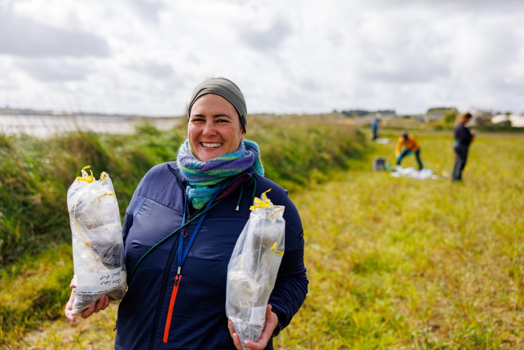
According to Arendt, this approach will also be crucial for the types of comparative studies his group is currently doing, in which they compare the nervous systems of many different animals to figure out what the brains of our distant ancestors looked like. For this purpose, the team will also be collecting organisms during the ongoing TREC expedition, to be analysed with volume electron microscopy and the new tools the scientists are developing.
And this is just the beginning for such applications. ”We used MoBIE to share a large number of tomograms of the SARS-CoV-2 virus, which COVID-19 researchers across the world have access to now,” said Schwab. The teams continue to collaborate with each other and across EMBL, making the dual tasks of knowledge-extraction and sharing from biological imaging data easier for the entire scientific community.
Funding
This research received additional support from various grants from the European Union’s Horizon 2020 research and innovation programme, the Chan Zuckerberg Initiative, and the European Research Council.
EMBL’s newest expedition attempts to answer some of the biggest questions in planetary biology, and will help scientists find solutions to pressing global concerns.