A remarkable new microscope built by EMBL researchers allows scientists to observe the dynamics of mechanical properties inside developing embryos in real time
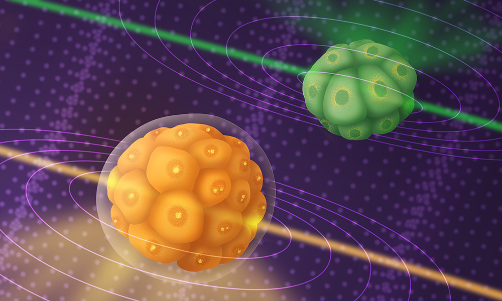
Line-scanning Brillouin microscopy (LSBM) – can be used to non-invasively study developing embryos in three dimensions and across time, providing novel biological insights. Credit: Joana Gomes Campos de Carvalho/EMBL
Summary
Scientists have come up with a new method to study the mechanical properties of developing embryos with unprecedented speed
The new method – line-scanning Brillouin microscopy (LSBM) – relies on a microscopy technique based on Brillouin scattering – a phenomenon where light interacts with naturally occurring thermal vibrations within materials.
In 1922, French physicist Léon Brillouin predicted an interesting phenomenon – when light is shone on a material, it interacts with the naturally occurring thermal vibrations within it, exchanging some energy in the process. This, in turn, influences how the light is scattered. By measuring the spectrum (colour) of the scattered light, we can deduce certain physical characteristics of that material.
More than a century later, scientists from the Prevedel group at EMBL, along with their collaborators, have harnessed this phenomenon (called Brillouin scattering) to track the mechanical properties of developing embryos with unprecedented speed and resolution.
“We often tend to think of cells or tissues only in terms of their biological properties – which genes do they express? Which biochemical pathways do they rely on? What chemical signals do they send to each other?” said Robert Prevedel, Group Leader at EMBL Heidelberg. “However, cells and tissues also have rich ‘mechanical’ lives. And these physical properties can help determine their biological function.”
The physical forces cells experience and their own material properties play a critical role in processes as diverse as embryonic development, tissue integrity, and the pathophysiology of diseases such as cancer. Two of these properties are viscosity – a measure of how easily a substance flows – and elasticity – a measure of how quickly a deformed object returns to its original state.
However, measuring these properties without damaging the cells and tissues can be challenging, as measurement methods often involve invasive approaches like ‘poking’ or ‘probing’ the samples in different ways. Here, as researchers have previously shown, microscopy based on Brillouin scattering can be useful for non-invasively viewing and assessing tissue mechanics.
Unfortunately, traditional Brillouin microscopy suffers from a few drawbacks. First, the speed of imaging is slow, since the method relies on collecting information from a single point on the sample at a time. Second, many biological tissues and cells are highly sensitive to light, and as a result, the long light exposures required in Brillouin microscopy can harm or deter the very processes that scientists wish to study.
In their new study published in Nature Methods, Prevedel and his colleagues describe a new microscopy method based on Brillouin scattering, that deals with the above challenges in innovative ways. Researchers in the Prevedel group are experts in developing advanced optical imaging techniques and pushing the frontiers of deep tissue microscopy.
As Carlo Bevilacqua, PhD student in the Prevedel lab and first author of the study, explains, the new method, called line-scanning Brillouin microscopy (LSBM), provides three major advantages.
First, instead of collecting information from a single point at a time, the new microscope scans the sample using an entire line of light at a time, which increases the speed of imaging by at least a hundred times. Second, it uses a new optical geometry, near-infrared light, and a Rubidium cell to significantly increase the signal-to-noise ratio, providing better resolution and reducing the risk of light-induced damage to cells.
And third, by integrating this system with an advanced light-sheet microscope, scientists can simultaneously visualise biomolecules using fluorescence, and this information can be layered on top of the mechanical properties of tissues.
“I really like building things,” said Bevilacqua. “Setting up a microscope like this involves quite a bit of theoretical optics knowledge, coupled with engineering and hands-on work, but you also require biological know-how.”
As proof of principle, the scientists used their newly built microscope to study embryonic development in three animal species from different evolutionary branches of the tree of life – fruit flies, mice, and a marine organism called Phallusia mammillata. In all of these species, the new microscopy method allowed researchers to follow the dynamics of mechanical changes in developing embryos in three dimensions and over a time scale of many hours.
“Studying development like this, in real time and in the whole volume of tissues rather than just the surface, can reveal many new and interesting biological mechanisms,” said Juan Manuel Gomez, postdoc in the Leptin and Prevedel labs and second author of the study.
The study involved collaborations with multiple other groups at EMBL, including the Ellenberg and Diz-Muñoz groups, as well as EMBL alumna Maria Leptin, formerly the director of EMBO and currently the European Research Council (ERC) President.
This method was also selected as one of The Guardian’s 10 biggest science stories of 2022. “That was a big surprise; I definitely didn’t see it coming,” said Prevedel. “I think it really shows the potential of the method, and the growing recognition of the importance of understanding how the biomechanical properties of tissues influence their function.”
Fare luce sulla meccanica dello sviluppo embrionale
Sintesi
Un giovane ricercatore italiano tra gli scienziati che hanno messo a punto un nuovo metodo per studiare le proprietà meccaniche degli embrioni in via di sviluppo con una velocità senza precedenti.
il nuovo metodo – la microscopia Brillouin a scansione di linea (LSBM) – consente ai ricercatori di seguire in modo non invasivo la dinamica dei cambiamenti meccanici nei tessuti in tre dimensioni e su una scala temporale di molte ore.
Nel 1922, il fisico francese Léon Brillouin predisse un fenomeno interessante: quando la luce viene irradiata su un materiale, interagisce con le vibrazioni termiche naturalmente presenti al suo interno, scambiando una certa energia nel processo. Questo, a sua volta, influenza il modo in cui la luce viene diffusa. Misurando lo spettro (colore) della luce diffusa, possiamo dedurre alcune caratteristiche fisiche del materiale.
Più di un secolo dopo, gli scienziati del gruppo di Robert Prevedel all’EMBL di Heidelberg, insieme ai loro collaboratori, hanno pubblicato uno studio su Nature Methods che sfrutta questo fenomeno (chiamato diffusione Brillouin) per tracciare le proprietà meccaniche degli embrioni in via di sviluppo con una velocità e una risoluzione senza precedenti. Primo autore della pubblicazione è Carlo Bevilacqua, dottorando italiano nel gruppo di Prevedel.
“Spesso tendiamo a pensare alle cellule o ai tessuti solo in termini di proprietà biologiche: quali geni esprimono? Su quali vie biochimiche si basano? Quali segnali chimici si inviano l’un l’altro?”, ha dichiarato Robert Prevedel, Group Leader dell’EMBL di Heidelberg. “Tuttavia, le cellule e i tessuti hanno delle proprietà fisiche che possono contribuire a determinarne la funzione biologica”.
Le forze fisiche che le cellule subiscono e le loro proprietà materiali giocano un ruolo fondamentale in processi diversi come lo sviluppo embrionale, l’integrità dei tessuti e la fisiopatologia di malattie come il cancro. Due di queste proprietà sono la viscosità – una misura della facilità di scorrimento di una sostanza – e l’elasticità – una misura della rapidità con cui un oggetto deformato ritorna al suo stato originale.
Tuttavia, misurare queste proprietà senza danneggiare le cellule e i tessuti può essere complicato, in quanto i metodi di misurazione spesso comportano approcci invasivi. In questo caso, come i ricercatori hanno precedentemente dimostrato, la microscopia basata sulla diffusione Brillouin può essere utile per visualizzare e valutare in modo non invasivo la meccanica dei tessuti.
La microscopia Brillouin tradizionale risente di alcuni inconvenienti. In primo luogo, la velocità di acquisizione delle immagini è lenta, poiché il metodo si basa sulla raccolta di informazioni da un singolo punto del campione alla volta. In secondo luogo, molti tessuti e cellule biologiche sono altamente sensibili alla luce e, di conseguenza, le lunghe esposizioni alla luce richieste dalla microscopia Brillouin possono danneggiare o alterare proprio i processi che gli scienziati desiderano studiare.
Nel nuovo studio pubblicato su Nature Methods, Prevedel e i suoi colleghi descrivono un nuovo metodo di microscopia basato sulla diffusione Brillouin, che affronta le suddette sfide in modo innovativo. I ricercatori del gruppo di Prevedel sono esperti nello sviluppo di tecniche avanzate di imaging ottico e nello spingersi oltre le frontiere della microscopia dei tessuti profondi.
Come spiega Carlo Bevilacqua, il nuovo metodo chiamato microscopia Brillouin a scansione di linea (LSBM), offre tre vantaggi principali.
Innanzitutto, invece di raccogliere informazioni da un singolo punto alla volta, il nuovo microscopio scansiona il campione utilizzando un’intera linea di luce alla volta, aumentando la velocità di imaging di almeno cento volte. In secondo luogo, utilizza una nuova geometria ottica, la luce del vicino infrarosso e una cella al rubidio per aumentare significativamente il rapporto segnale/rumore, fornendo una migliore risoluzione e riducendo il rischio di danni alle cellule indotti dalla luce.
In terzo luogo, integrando questo sistema con un microscopio avanzato a fogli luminosi, gli scienziati possono visualizzare simultaneamente le biomolecole utilizzando la fluorescenza, e queste informazioni possono essere sovrapposte alle proprietà meccaniche dei tessuti.
“Mi piace molto costruire nuovi strumenti”, ha detto Bevilacqua. “La messa a punto di un microscopio come questo implica una certa conoscenza dell’ottica teorica, unita all’ingegneria e al lavoro pratico, ma è necessario anche un know-how biologico”.
Gli scienziati hanno utilizzato il microscopio appena costruito per studiare lo sviluppo embrionale in tre specie animali appartenenti a diversi rami evolutivi dell’albero della vita: moscerini della frutta, topi e un organismo marino chiamato Phallusia mammillata. In tutte queste specie, il nuovo metodo di microscopia ha permesso ai ricercatori di seguire la dinamica dei cambiamenti meccanici negli embrioni in via di sviluppo in tre dimensioni e su una scala temporale di molte ore.
“Studiare lo sviluppo in questo modo, in tempo reale e nell’intero volume dei tessuti piuttosto che solo sulla superficie, può rivelare molti nuovi e interessanti meccanismi biologici”, ha dichiarato Juan Manuel Gomez, postdoc nel laboratorio di Leptin e Prevedel e secondo autore dello studio.
Lo studio ha visto la collaborazione di molti altri gruppi dell’EMBL, tra cui i gruppi Ellenberg e Diz-Muñoz, nonché dell’ex ricercatrice dell’EMBL Maria Leptin, già direttore dell’EMBO e attualmente presidente del Consiglio Europeo della Ricerca (ERC).
Questo metodo è stato anche selezionato dal ‘The Guardian’ come una delle 10 ricerche scientifiche più importanti del 2022. “È stata una grande sorpresa, non me l’aspettavo”, ha dichiarato Prevedel. “Penso che dimostri davvero il potenziale del metodo e il crescente riconoscimento dell’importanza di capire come le proprietà biomeccaniche dei tessuti influenzino la loro funzione”.