Spotlight: Hot waters, shrinking fish
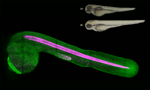
EMBL scientists investigate how zebrafish bodies change when grown at higher temperatures.
SCIENCE & TECHNOLOGY2025
science-technology
Showing results out of
EMBL scientists investigate how zebrafish bodies change when grown at higher temperatures.
SCIENCE & TECHNOLOGY2025
science-technology

Jia Le Lim talks about contributing back to society through science, finding inspiration in the way birds fly in the sky, and winning a recent international piano competition.
PEOPLE & PERSPECTIVES2024
people-perspectives
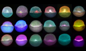
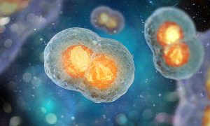
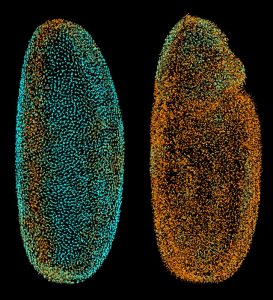
Zebrafish embryos during gastrulation, a very early stage of development, to study the effect of temperature on vertebrate embryo development.
LAB MATTERSSCIENCE & TECHNOLOGY2022
lab-matterspicture-of-the-weekscience-technology
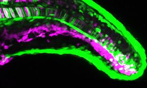
How do gene expression patterns result in the generation of different cell types? Scientists at EMBL Heidelberg used the zebrafish notochord to find out.
SCIENCE & TECHNOLOGY2022
sciencescience-technology

Michael Dorrity, one of EMBL’s newest group leaders, is studying how the environment influences early life stages in zebrafish.
LAB MATTERSPEOPLE & PERSPECTIVES2022
lab-matterspeople-perspectives

EMBL scientists have combined artificial intelligence (AI) algorithms with two cutting-edge microscopy techniques.
SCIENCE & TECHNOLOGY2021
sciencescience-technology
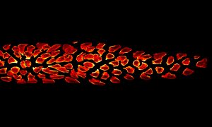
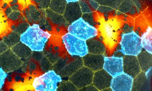
This group of cells represents an interesting example of organ formation where cells simultaneously move and change their shapes in a highly coordinated manner.
SCIENCE & TECHNOLOGY2020
picture-of-the-weekscience-technology
Researchers from the Sharpe group at EMBL Barcelona have published a method to track the developmental history of a cell using the gene editing tool CRISPR–Cas9, but without the need to create transgenic organisms.
SCIENCE & TECHNOLOGY2020
sciencescience-technology
In the Trivedi Group at EMBL Barcelona, Krisztina Arató and Jia Le Lim study the early development of zebrafish embryos.
SCIENCE & TECHNOLOGY2020
picture-of-the-weekscience-technology
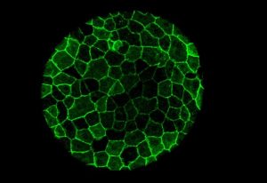
This beautiful mosaic of mostly hexagonal cells is the outer skin layer of a zebrafish larva as seen under a microscope. Each skin cell exhibits a unique pattern of actin ridges. Actin is a family of globular multifunctional proteins found in almost all eukaryotic cells. Actin forms microfilaments,…
SCIENCE & TECHNOLOGY2019
picture-of-the-weekscience-technology
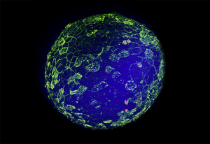
Despite missing the characteristic stripes one would expect from a zebra – or a zebrafish – the fractals in this Picture of the Week show a zebrafish; or at least some cells in a zebrafish embryo, a few hours after fertilisation. Zebrafish are not only popular aquarium fish, they are also an…
SCIENCE & TECHNOLOGY2019
picture-of-the-weekscience-technology

Migrating cells, it seems, cover their tracks not for fear of being followed, but to keep moving forward. Scientists at the European Molecular Biology Laboratory (EMBL) in Heidelberg, Germany, have now shown that cells in a zebrafish embryo determine which direction they move in by effectively…
SCIENCE & TECHNOLOGY2013
sciencescience-technology
The scientists at the European Molecular Biology Laboratory (EMBL) in Heidelberg, Germany, who ‘fathered’ the Digital Embryo have now given it wings, creating the Fly Digital Embryo. In work published today in Nature Methods, they were able to capture fruit fly development on film, and were the…
SCIENCE & TECHNOLOGY2010
sciencescience-technology
Researchers at the European Molecular Biology Laboratory (EMBL) have generated a digital zebrafish embryo – the first complete developmental blueprint of a vertebrate. With a newly developed microscope scientists could for the first time track all cells for the first 24 hours in the life of a…
SCIENCE & TECHNOLOGY2008
sciencescience-technology
No results found