
Accidental beauty
In nature, a fish embryo develops under the outer layer of the egg – the chorion. If all goes well, it grows strong until it breaks out of the egg into the world.
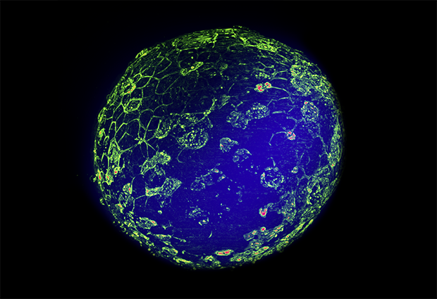
In the Trivedi Group at EMBL Barcelona, Krisztina Arató and Jia Le Lim study the early development of zebrafish embryos. As the structure of the embryo grows more complex, a contractile network made up of two proteins – actin and myosin – helps cells rearrange. To understand how this process works, the researchers must examine embryos that are stripped of their protective outer layer.
In this image, myosin appears in green on the embryo’s surface, labelled with green fluorescent protein (saturated pixels are in red), while the bulk of the embryo shows as a blue halo in a different focal plane. When Krisztina Arató saw this embryo after a night of live imaging, she realised something had gone wrong.
Despite standard sterilisation procedures, a microbial invader had managed to sneak into the medium and attack the embryo. Some embryonic cells had already undergone apoptosis – a form of programmed death that can occur in response to stress, for example during an infection. Here, large punctuated patches of myosin reveal dying cells.
The two researchers had to take a closer look to figure out what had happened. They captured this striking scene – resembling a satellite image of the Earth – with a multiview light-sheet microscope (MuVi-SPIM).

If you have a stunning picture of your science, your lab, or your site, you can submit it here.


