
Skin mosaic
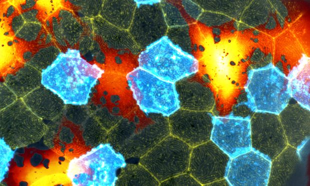
This beautiful mosaic of mostly hexagonal cells is the outer skin layer of a zebrafish larva as seen under a microscope.
Each skin cell exhibits a unique pattern of actin ridges. Actin is a family of globular multifunctional proteins found in almost all eukaryotic cells. Actin forms microfilaments, which are seen as yellow lines in the image. The ridges are thought to help with mucus retention, and could provide necessary plasticity and rigidity to surface cells. Whatever the reason for these ridges, their presence across many living species suggests they have an important function.
Eva Hasel, a postdoc in the Leptin group, captured this image during her research on the skin layers of zebrafish larvae and how skin cells respond to a type of inflammatory cell death. The zebrafish larva was imaged under the Zeiss 780 confocal microscope, using a 40x objective. The pattern of actin ridges is shown in yellow, and the mosaic membrane label is shown in cyan. The big underlying cells are melanocytes, which are false-coloured using a lookup table.

If you have a stunning picture of your science, your lab or your site, you can submit it to mathias.jaeger@embl.de.


