Molecular solar panels can help scientists control brain cells
Kirill Kovalev, an EMBL Hamburg researcher, is studying the structure of an ancient bacterial molecule to help scientists control brain cell activity
Issue 99
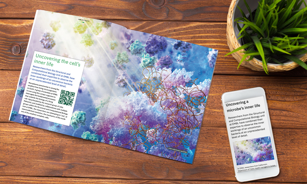
Researchers have observed the inner workings of an unusual bacteria at an unprecedented level of detail
In 1944, researchers from California’s Department of Public Health managed to isolate the causative agent for atypical pneumonia, a respiratory illness that afflicted many military personnel during World War II. First called the ‘Eaton Agent’ after the researcher who isolated it, the pathogen was believed for many years to be an unidentified virus. It was not until the 1960s that scientists determined conclusively that the infectious agent causing atypical pneumonia was not a virus, but a very small bacterium.
Mycoplasma pneumoniae, as the species was later named, is among the tiniest free-living microorganisms on Earth. Its genome contains only 687 genes. For comparison, Escherichia coli, the bacteria most commonly used in lab studies, has over 4,400 genes, while human beings have over 20,000. At less than 1 micron in length, 1.5 trillion M. pneumoniae could fit into a single droplet of water. When it infects humans, it parasitises respiratory tract cells, often evading the immune system during the process. Also, unusual for bacteria, it lacks a cell wall surrounding its plasma membrane.
A group of EMBL scientists is now using this unique organism to interrogate fundamental processes that underlie the biology of a living cell. Spearheaded by the research group of Julia Mahamid, and enabled by collaborations with multiple groups within and outside EMBL, the researchers are attempting to ‘see’, at unprecedented resolution, the mechanisms of life inside one of the smallest living cells.
Researchers from EMBL’s Structural and Computational Biology unit established Mycoplasma pneumoniae as a model organism in the early 2000s. Teams led by Peer Bork, Luis Serrano, and Anne-Claude Gavin sequenced and annotated its genome, which was followed by the publication of three seminal papers delineating its metabolomics, proteomics, and transcriptomics, respectively.
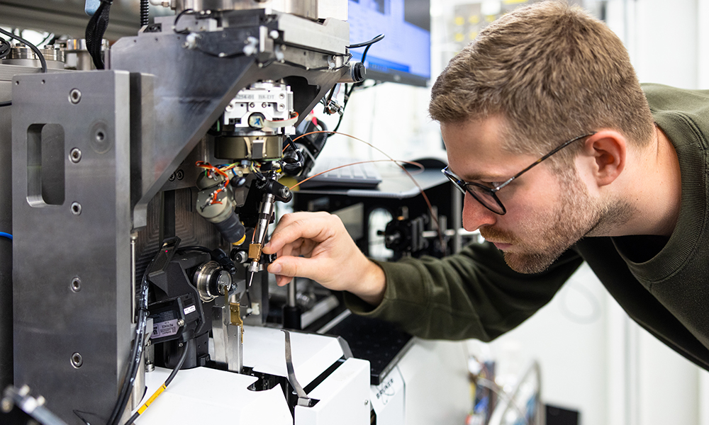
Two decades earlier, Jacques Dubochet, another EMBL researcher, had developed a way to prepare and image biological samples using cryo-electron microscopy (cryo-EM), work that won him the Nobel Prize in 2017. Relying on a method of freezing biological samples extremely quickly to prevent the formation of ice crystals, cryo-EM allows researchers to observe the structure of complex biomolecules at atomic resolution.
Soon afterwards, researchers developed the technique of cryo-electron tomography (cryo-ET), another big leap in harnessing the power of electron microscopy. While cryo-EM allows scientists to observe the structure of biological molecules, it usually requires samples of carefully isolated molecules taken out of their cellular context. However, cryo-ET allows scientists to take snapshots of intact cells, along with all their internal components, which can later be reconstructed in 3D.
Given Mycoplasma’s small size, it is possible to obtain a series of images to combine into a clear picture of the entire cell and all its constituents. When Mahamid started her group at EMBL in 2017, she decided to harness the growing power and resolution of advanced cryo-ET technologies to study cellular mechanisms in action inside this model organism.
“Personally, I’m amazed by the ability to do structural biology inside cells,” said Joe Dobbs, one of the PhD students in the Mahamid group. “Cryo-EM is well-known for its ability to provide insight into the structures of macromolecular complexes in purified samples, but actually seeing the insides of cells, and how molecules interact with each other in context, is incredible.”
The study has attracted a diverse group of scientists and engineers. Vastly differing in areas of interest and expertise, they are united by their belief in the potential for M. pneumoniae to serve as a window into the life of a cell.
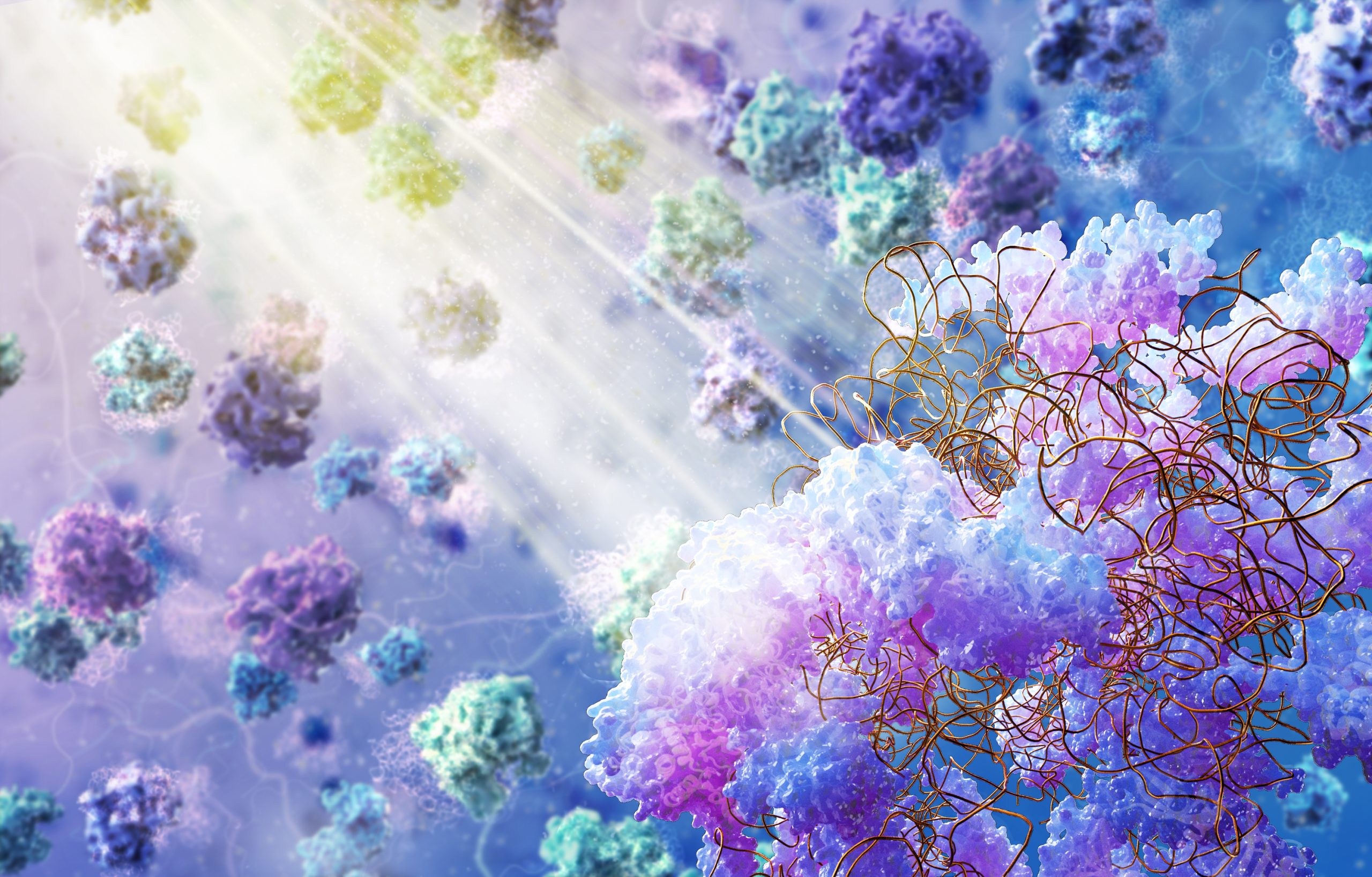
For example, Judith Zaugg started her lab at EMBL in 2014 and is interested in understanding the molecular basis of complex genetic traits and diseases, for which she usually works with human genomics data. With Mycoplasma, she became fascinated by the idea of observing gene expression in action within a living cell. In the cryo-ET images that the Mahamid group grabs, DNA can be observed as filamentous structures and the RNA polymerase enzymes seen attached to them, and Zaugg believes that one day this might be used to figure out which regions of DNA or genes are active – or being ‘transcribed’ – at a given point in time.
“It is very exciting to ‘see’ transcription in progress,” said Zaugg. “With this model, we can perhaps one day address very fundamental biological questions like – how does the cell reorganise its DNA upon receiving a certain stimulus? While we know (based on genomics data) that genes are differentially expressed based on stimuli, we still don’t know how the cell rearranges its internal structure and genome to achieve this.”
With future advances in correlative electron and fluorescence microscopy, Zaugg hopes that answering questions like these might become possible. In turn, her team brings to the project computational expertise to mine the data, which otherwise could have taken months or even years to process.
Peer Bork, who co-initiated and coordinated the initial proteomic, transcriptomic, and metabolomic characterisation of M. pneumoniae in the 2000s, is interested not only in the infectious disease aspects of this model system but also in its potential for understanding molecular networks. His group has aided the study with computational expertise as well as by connecting global bioinformatic data.
”This project leverages some of EMBL’s key strengths – collaborating and coordinating across a diversity of disciplines to answer fundamental biological questions that cannot be addressed otherwise,” said Bork.
Maria Zimmermann-Kogadeeva, another EMBL group leader, started collaborating with Mahamid while still a postdoc in the Bork group. Her computational insights played a key role in a recent project that studied the machinery of protein synthesis in action inside the cell. She is also involved in a project focused on studying previously uncharacterised membrane proteins that can be observed in cryo-ET images of Mycoplasma.
“The cryo-ET technology allows us to look deep inside the cell, and understand how intracellular processes change under different conditions,” said Zimmermann-Kogadeeva. “The data obtained is of very high quality and can be combined with other molecular datasets to ask interesting questions.” She and Bork are both optimistic about the potential of investigating cellular metabolomics within Mycoplasma in the future.
“Our goal is to connect biology across scales,” said Mahamid. “Mycoplasma gives us a way to study biological systems all the way across from the nanometre range of molecules to the micrometre range of entire cells.”
While cryo-ET allows scientists to look at the whole cell, it doesn’t make it easy to identify what they are seeing. Unlike with fluorescence microscopy, it isn’t easy to tag or label specific objects inside the cell and distinguish them from their neighbours in cryo-ET. As a result, scientists are effectively left with an incomplete map, most of which has no helpful labels.
Soheil Mojiri, a postdoctoral fellow in the Ries group, is building a microscope to solve this problem. An engineer by training, Mojiri is fascinated by the possibility of exploiting optical physics to address challenging biological questions. His goal is to fruitfully marry cryo-ET and super-resolution optical microscopy at low temperatures.
“Cryo-ET allows us to look at molecular architecture and molecules in context, but we’re limited by what we can identify in the extremely noisy, crowded, and heterogeneous cellular environment. So while we can just about make out big things, such as ribosomes – the cell’s protein production machines, smaller molecules and complexes can escape our notice unless we have prior information to use,” said Mojiri.
Mojiri is developing a prototype microscope that would allow scientists to take a frozen cell sample, visualise individual proteins or complexes with fluorescent tags that are genetically introduced into live cells, and later subject the same sample to cryo-ET. Comparing the images obtained by the two methods would let researchers leverage electron microscopy’s structural resolution as well as fluorescence microscopy’s specificity.
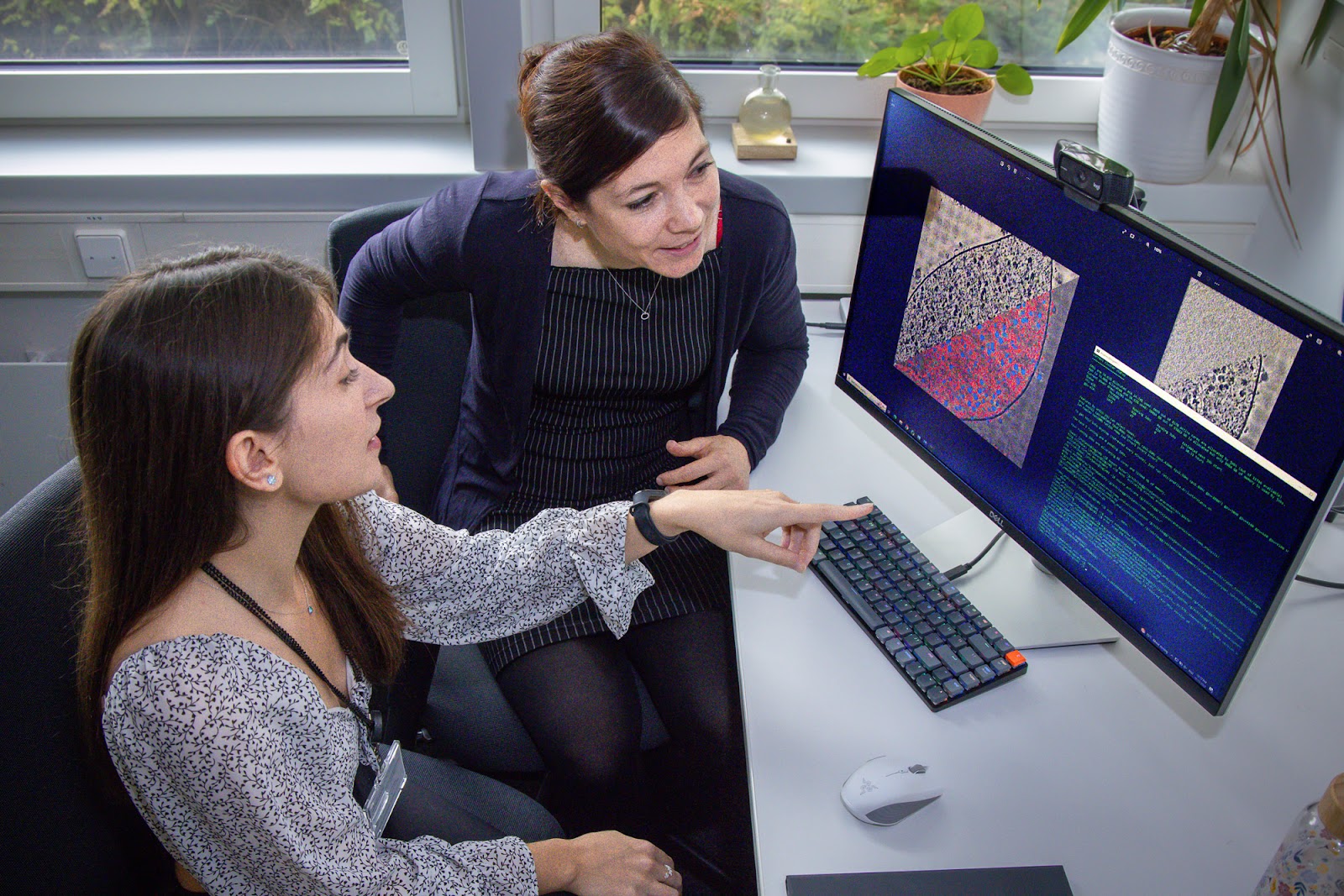
Not all technology development for this project is on the hardware side, however. The researchers have generated a huge volume of data, which presents challenges in analysing it to draw new insights. This is where Anna Kreshuk, with her expertise in machine-learning-based image analysis, steps in. Kreshuk and her team create innovative AI-based methods that find meaningful information from biological images, often gigabytes in size and containing a lot of background information that may not be relevant.
Ricardo Sanchez, an ARISE fellow at EMBL Heidelberg, has been working on finding ways around this challenge. “With cryo-ET, one of the biggest problems is reducing the signal-to-noise ratio and identifying structures of interest among all the cellular background,” he said. “My goal is to solve this problem with the least amount of computational resources.”
“What fascinates me is the completeness of this system,” said Kreshuk. “It’s the whole living thing. Everything that it needs to be an independent (and pretty dangerous) living organism is visible in that one image.”

The project is already yielding dividends when it comes to understanding fundamental biological processes that make life possible. In a recently published paper, Liang Xue from the Mahamid group led a study that allowed the team to visualise the process of translation – the synthesis of new proteins – inside the cell at atomic detail. The structures they focused on is probably one of the easiest to identify in a cryo-ET image – ribosomes. Ribosomes are some of the most ancient molecular machines present in all living organisms and essential for protein synthesis.
“Ribosomes not only play an essential role in genetic information flow but also serve as platforms to monitor cellular states, e.g. cell stress response,” said Xue. “Inside living cells, ribosomes function as highly interconnected networks of molecular machines.”
Using high-quality cryo-ET Mycoplasma data, the researchers were not only able to observe the dynamic structural changes that took place in ribosomes as they proceeded through the protein synthesis cycle, but they could also observe what happens to these processes when antibiotics perturb cells. Since the translation machinery is quite similar in structure and function throughout the tree of life, the study can be extrapolated to other prokaryotic or eukaryotic species that are more challenging to image with cryo-ET.
“I hope my work at EMBL establishes a framework to do high-resolution structural biology inside the cell,” said Xue. “Ribosomes and Mycoplasma are just the beginning. With more and more molecular machines resolved inside cells, we are heading toward the ultimate goal of building an atomic cell model.”
The researchers are also using Mycoplasma to understand the role of unknown membrane proteins that can be observed in cryo-ET images In these studies, Jan Kosinski’s and Christian Löw’s groups at EMBL Hamburg and Nassos Typas’s group at EMBL Heidelberg are also involved. A tool they have turned to often in these investigations is AlphaFold, the AI algorithm that predicts protein structures, whose developers won the 2023 Breakthrough Prize in life sciences.
The project also involves collaborations with scientists outside EMBL and in EMBL member states, including the groups of Jörg Stülke, Georg-August-Universität Göttingen, Patrick Cramer, Max Planck Institute for Multidisciplinary Sciences, Juri Rappsilber, Institut für Biotechnologie, Technische Universität Berlin, and Luis Serrano, Centre for Genomic Regulation, Barcelona.
While research in the last few decades has helped to rapidly identify and annotate many species’ genomes, our knowledge of the function and precise roles of most genes and proteins remains incomplete, even for an organism as simple as Mycoplasma. Projects like these are a key step towards bridging this knowledge gap. EMBL’s new programme ‘Molecules to Ecosystems’ aims to study living organisms in the context of their environment. In doing so, scientists will doubtless encounter many more new molecules and biological processes, which this project paves the way towards studying and understanding in detail. It also exemplifies the collaborative approaches at the heart of this new programme.
“I think it is a pretty long and hard way from imaging Mycoplasma to the data interpretation. We need diverse expertise and knowledge to pave this way,” said Mojiri. “In particular, we require biologists, physicists, and data scientists.” The collaborative atmosphere at EMBL has made such a coming together of talents and knowledge possible. The team gathers in a monthly meeting dedicated to the Mycoplasma project to exchange ideas, discuss the progress of experiments, and troubleshoot challenges.
“Mycoplasma has turned out to be a powerful model for studying the dynamics of molecular machines and understanding intracellular cross-talk,” said Mahamid. “The collaborative approach was absolutely crucial to this. To realise the massive potential of cryo-ET and the Mycoplasma model, we will need to continue as we have started, together and moving forward.”
Kirill Kovalev, an EMBL Hamburg researcher, is studying the structure of an ancient bacterial molecule to help scientists control brain cell activity

Newts act as model organisms for Maria Tosches, winner of the 2022 John Kendrew Award, to further explore the cellular makeup of vertebrate brains.
Click here to download a quick overview of all the articles included in this digital issue of EMBLetc.