
Alba Diz-Muñoz
Interim Head of Unit for CBB
ORCID: 0000-0001-6864-8901
EditUnderstanding how the fundamental unit of life, the cell, functions molecularly and physically
In this unit, physicists, chemists and biologists work closely together to elucidate the fundamental rules that govern dynamic cell organisation and function. Groups are developing new instruments and technologies to reach this ambitious goal.
Cells are the smallest autonomous units of life and occupy the midpoint between the molecular and macroscopic scales. In order to understand how living systems are built and function, we need to understand the physical principles that underlie cellular organisation and function.
It is in the cell where we will first understand the basic processes of life at the molecular level in a physiological context. The cell provides the natural coordinate system in space and time onto which we have to map and integrate genomic, transcriptomic, proteomic, structural and biophysical information about the molecules that make up living systems. In short, cell biology has become an integrative hub of much of modern biological research.
This is a time of tremendous opportunity for cell biology, but realising it also represents a formidable challenge and requires new concepts and approaches. Individual cellular processes ─ such as signalling, membrane trafficking, cytoskeletal dynamics and cell migration, gene expression or cell division ─ can no longer be studied in isolation but need to be considered as integrated events. The default situation is that the molecular machinery that performs these functions is complex and combinatorial at the single protein, protein complex, and pathway level. This requires new ways of thinking about cellular functions that use network biology and employing quantitative theoretical methods to generate mechanistic and predictive models that rely on realistic physical principles at the cellular, subcellular and molecular scale. Therefore, cell biology needs to integrate traditionally separate disciplines to realise its potential.
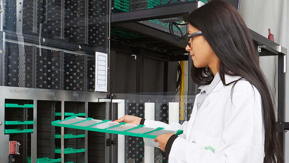
Novel developments in microscopy, computer simulations and chemical biology-based probes are a particular strength of the Unit. We constantly explore new directions and integrate new approaches and disciplines to answer cell biological questions. New correlative light/electron and super-resolution imaging methods, as well as mechanistic biochemistry, allow us to directly interface between cell and structural biology to understand molecular mechanisms. Furthermore, advances in live and deep tissue imaging methods now allow us to carry out cell biology in developing organisms to understand how collective cell behaviour leads to organ formation, and how cells interact with their physiological microenvironment.
Mechanisms of cellular functions are often best understood when the organisation of the cell changes dramatically to carry out new functions. This is the case when cells divide, when they change their fate. Both opportunities are exploited in the Unit. As a cell prepares to divide, all the microtubules suddenly depolymerise to reassemble into the mitotic spindle. At the same time, the nucleus is disassembled, mitotic chromosomes are formed, the Golgi complex fragments and membrane traffic ceases. After segregation of the genome is achieved, cellular organisation is re-established. Thus every cell cycle provides the opportunity to study the principles of the biogenesis of cellular compartments. Similarly, the genetic programme is changed and a reorganisation of cellular architecture takes place, guided by rules that we begin to unravel when progenitor cells differentiate into new cell types or start to migrate. Understanding these rules and principles is our challenge in the years to come.
Interim Head of Unit for CBB
ORCID: 0000-0001-6864-8901
Edit
Interim Head of Unit for CBB
ORCID: 0000-0003-1334-6388
Edit
Understanding cross-scale principles of cellular organisation
Edit
Cellular phase separation by surfactants
Edit
Building next-generation fluorescent tools for biological imaging
Edit
Mechanobiology at the cell surface
Edit
Cell division and nuclear organisation
Edit
Theory of cellular and multicellular organisation
Edit
Self-organisation in meiosis
Edit
Machine learning for bioimage analysis
Edit
Membrane traffic and organelle biogenesis
Edit
Advanced optical techniques for deep tissue microscopy
Edit
Principles of genome self-organisation
Edit
Volume correlative light and electron microscopy
Edit
Advanced Light Microscopy technology development and service provision
Edit
Regulation of stem cell fate
Technological developments in microscopy have made cell biology a quantitative and data intensive discipline with many computational challenges
The Euro-BioImaging Bio-Hub is part of the Euro-BioImaging ERIC headquarter and is hosted by EMBL in Heidelberg
Unit Administrator
Edit
Scientific Officer
EditResearch Scientist in Lipidomics, Visiting Associate Professor from SDU
EditUnit Administrator
Edit
Lab Manager
Edit


Advancing molecular biology research to study life in context

Research groups at EMBL are organised into nine units spanning six European sites

Explore our latest vacancies and sign up for job alerts to get notified when something suitable comes up