Cell map unlocks secrets of how reproductive organs form
A new single-cell map reveals how human reproductive organs form and respond to environmental influences
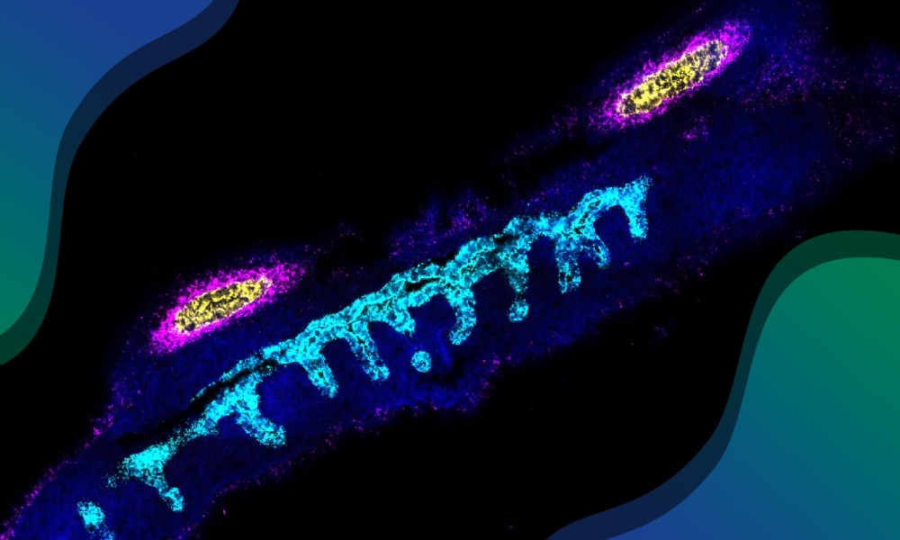
A developing human reproductive tract stained for marker of the Wolffian duct (cyan) and markers of the regressing Mullerian Duct (yellow and magenta). Credit: Cecilia Icoresi Mazzeo, Staff Scientist/Wellcome Sanger Institute
New research has mapped the cell types that specialise to form reproductive organs in both sexes, identifying key genes and signals that drive this process. The findings offer important insights into conditions affecting the reproductive organs and how environmental chemicals may affect reproductive health.
Researchers at EMBL’s European Bioinformatics Institute (EMBL-EBI) and the Wellcome Sanger Institute used a combination of single-cell and spatial genomics technologies to analyse over half a million human cells from the developing reproductive system.
This study, published in the journal Nature, provides the most detailed picture to date of how reproductive organs form in the womb, uncovering crucial biological pathways that shape their development.
“For the first time, we can see in detail how the human reproductive system is assembled before birth,” said Valentina Lorenzi, Staff Scientist at the Wellcome Sanger Institute and former PhD student at EMBL-EBI in the Marioni Group. “This map pinpoints the exact cells and signals that shape each organ, and highlights when development is most vulnerable. This is an essential step towards understanding fertility, congenital alterations, and the impact of our environment on reproductive health.”
How does human sex differentiation occur?
Although chromosomal sex (XX or XY) is determined at conception, visible differences in the developing reproductive system do not appear until about six weeks later. At this stage, all embryos have undifferentiated gonads as well as the same paired structures – the Müllerian ducts and the Wolffian ducts – which have the potential to form the female or male internal reproductive organs.
A gene on the Y chromosome called SRY triggers the undifferentiated gonads to develop into the testes; the testes then produce hormones that guide the Wolffian ducts to form male reproductive structures and cause the Müllerian ducts to regress. In the absence of SRY, the gonads become ovaries, and the Müllerian ducts develop into female reproductive organs.
Mapping reproductive development
While development of the gonads has been mapped at the cellular and molecular level in detail due to their importance in fertility, the development of the rest of the reproductive system has been far less understood, until now.
In this study, researchers analysed over half a million individual cells from 89 donated embryonic and fetal tissue samples, to look at what genes and signals underpin the differentiation of the male and female reproductive tract. The study is part of the Human Cell Atlas initiative, which is mapping all cell types in the body to understand health and disease.
“Many conditions affecting fertility and reproductive health have their roots in development before birth, yet until now we lacked a full picture of how these organs form in humans,” said Roser Vento-Tormo, Group leader at the Wellcome Sanger Institute. “Our atlas offers that missing piece, providing a powerful resource for both basic science and clinical research.”