14 May 2026

A newly identified protein, SNOR, has been found to help cells in a starvation-induced dormancy restart protein synthesis once nutrients are again available.
SCIENCE & TECHNOLOGY
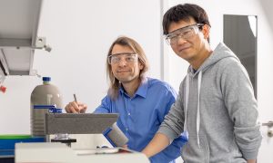
28 January 2026
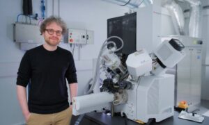
The new EMBL Grenoble group leader will explore the origin of eukaryotes and their complex cellular organisation by studying Asgard archaea and other non-model microorganisms.
PEOPLE & PERSPECTIVES
3 January 2025
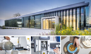
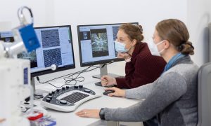
Scientists share their experiences of creating stunning images at the EMBL Imaging Centre.
CONNECTIONS
17 October 2024
Imaging lets us observe biology in action – it makes visible the hidden processes of life. From its founding, EMBL has been a centre of breakthroughs and developments in bioimaging, and it continues to play a pioneering role in this field today.
SCIENCE & TECHNOLOGY
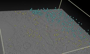
8 October 2024
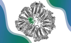
Scientists at EMBL Heidelberg and University of Virginia revealed a new cellular response to starvation: ribosomes attach to the mitochondrial outer membrane in a very unusual way, via their small subunit. The finding made in yeast might provide insights into how cancer cells survive the harsh…
SCIENCE & TECHNOLOGY
10 April 2024
Data obtained through cryogenic sample electron tomography (cryo-ET) are openly available in the new Electron Microscopy Public Image Archive (EMPIAR) cryo-ET tomography browser.
EMBL ANNOUNCEMENTS
2024
announcementsembl-announcements
3 April 2023
Home to some of Europe’s most cutting-edge tools in molecular biology, EMBL has long shared its expertise and access to these tools through an extensive repertoire of courses, conferences, seminars, and other training. And now included in this mix is a job shadowing programme at EMBL Imaging…
LAB MATTERSSCIENCE & TECHNOLOGY
2023
lab-mattersscience-technology
20 March 2023
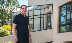
Together with his team, Gergely Papp pushes the frontiers of technology development in the field of structural biology.
LAB MATTERSPEOPLE & PERSPECTIVES
2023
lab-matterspeople-perspectives
28 September 2022
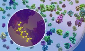
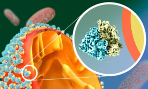
New research by EMBL scientists shows at atomic detail how antibiotics affect the process of protein production inside bacteria.
SCIENCE & TECHNOLOGY
2022
sciencescience-technology
10 June 2022
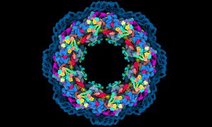
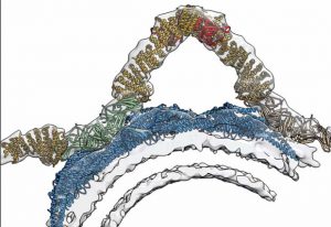
Scientists have solved several mysteries around the structure and function of a true molecular giant: the human nuclear pore complex. They created the most complete model of the complex thanks to combining the program AlphaFold2 with cryo-electron tomography, integrative modelling, molecular…
SCIENCE & TECHNOLOGY
2022
sciencescience-technology
21 December 2021
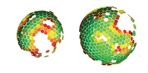
EMBL Hamburg’s Kosinski Group, the Beck Laboratory at the Max Planck Institute of Biophysics, and colleagues at EMBL Heidelberg recorded the nuclear pore complex contracting in living cells. They visualised the movement with an unprecedented level of detail with help of new software called…
SCIENCE & TECHNOLOGY
2021
sciencescience-technology
12 November 2021
Correlative microscopy service enables PhD student from Switzerland to study structure and location of proteins cells use to communicate.
LAB MATTERSSCIENCE & TECHNOLOGY
2021
lab-mattersscience-technology
20 April 2021
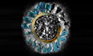
What does coronavirus’s spike protein look like in 3D? EMBL scientists and colleagues used cryo-electron tomography and molecular dynamics simulations to find out.
SCIENCE & TECHNOLOGY
2021
picture-of-the-weekscience-technology
23 February 2021
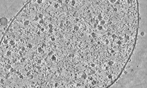
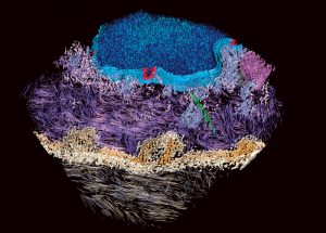
Liang Xue used cryo-electron tomography to capture this detailed image of a Mycoplasma pneumoniae cell.
SCIENCE & TECHNOLOGY
2021
picture-of-the-weekscience-technology
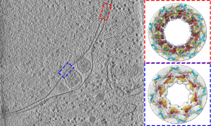
7 December 2020

While cryo-electron tomography (cryo-ET) was first envisioned in 1968, the advances the Mahamid group are bringing to this 3D method for studying molecules directly inside cells are new, and are likely to greatly expand its use.
SCIENCE & TECHNOLOGY
2020
sciencescience-technology
3 September 2020
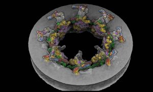
Scientists from the Beck group have studied the 3D structure of nuclear pores in budding yeast. They show how the architecture of the nuclear pore complex differs inside cells compared to its form observed in vitro studies.
SCIENCE & TECHNOLOGY
2020
sciencescience-technology
31 July 2020
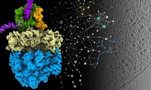
A new approach that allows researchers to see molecular machinery at work inside cells has offered a deeper understanding of how bacteria produce proteins and a unique glimpse into how they respond to antibiotics.
SCIENCE & TECHNOLOGY
2020
sciencescience-technology
17 April 2020

EMBL Heidelberg reopens the Cryo-Electron Microscopy Service Platform to support coronavirus structural biology research.
CONNECTIONS
18 November 2019
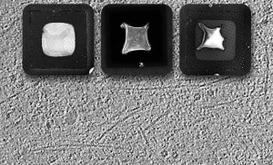
A new technique in cryo-EM
SCIENCE & TECHNOLOGY
2019
sciencescience-technology
17 September 2018
Retromer’s 3D structure improves understanding of cellular sorting and packaging
SCIENCE & TECHNOLOGY
2018
sciencescience-technology
23 June 2009
Scientists at the European Molecular Biology Laboratory (EMBL) and the University Clinic Heidelberg, Germany, have produced a three-dimensional reconstruction of HIV (Human Immunodeficiency Virus), which shows the structure of the immature form of the virus at unprecedented detail. Immature HIV is…
SCIENCE & TECHNOLOGY
2009
sciencescience-technology
5 December 2007
Seeing proteins in their natural environment and interactions inside cells has been a longstanding goal. Using an advanced microscopy technique called cryo-electron tomography, researchers from the European Molecular Biology Laboratory (EMBL) have visualised proteins responsible for cell-cell…
SCIENCE & TECHNOLOGY
2007
sciencescience-technology
No results found