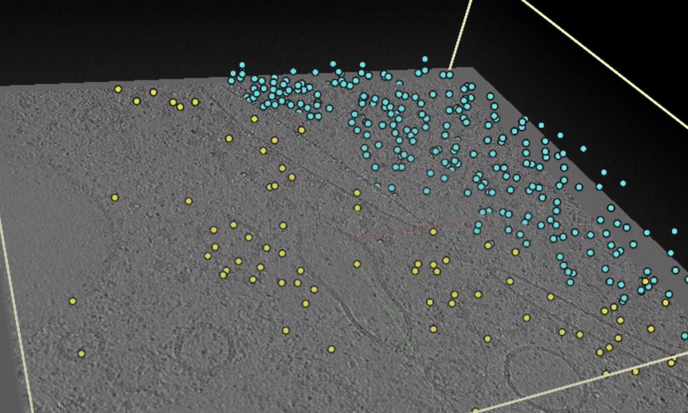
Exploring cellular complexity with EMPIAR’s new cryo-electron tomography prototype browser
Data obtained through cryogenic sample electron tomography (cryo-ET) are openly available in the new Electron Microscopy Public Image Archive (EMPIAR) cryo-ET tomography browser
Over 1,800 reconstructed cryo-ET tomograms and associated raw data of Chlamydomonas reinhardtii are freely available in the EMPIAR database
A selected subset of these image data are accessible, visualisable, and easily navigable using EMPIAR’s new tomogram browser
Cryo-ET allows scientists to visualise proteins in 3D in their natural environment, bridging the gap between structural and cellular biology and laying the groundwork for advancements in disease treatment, drug development, and more.
Data obtained through cryogenic electron tomography (cryo-ET), a cutting-edge technology that offers the highest resolution images of cellular structures, will be made openly available in the new Electron Microscopy Public Image Archive (EMPIAR) cryo-ET browser.
This new dataset, provided by Thermo Fisher Scientific with work from other research colleagues, showcases the power of cryo-ET in revealing the intricate details of the cell at individual protein resolution and lays the foundation for advancements in the field of structural biology.
“These cryo-ET data are a leap forward in imaging technology. They provide a transformative shift in how we understand cellular functions and the behaviour of proteins in their natural environments,” said Sameer Velankar, Team Leader and Senior Scientist at EMBL-EBI. “This collection is an invaluable resource for the scientific community that will help enrich our understanding of cellular mechanisms.”
What is cryo-ET?
Cryo-ET involves flash-freezing biological specimens to capture them in their natural state, avoiding the distortion or damage that traditional preparation methods might cause. This technique then uses a beam of electrons to image the sample, creating a series of two-dimensional images from different angles. These images are reconstructed into detailed three-dimensional models, offering insights into the architecture of cells at the level of individual proteins without the need for crystallisation, allowing scientists to visualise proteins in their natural state and cellular context.
Exploring the microscopic universe
Cryo-ET allows scientists to visualise proteins in their cellular environment in 3D, bridging the gap between structural and cellular biology. Today, over 1,800 reconstructed tomograms and associated raw data of Chlamydomonas reinhardtii have been made freely available in the EMPIAR database helping researchers to investigate the mechanisms behind cellular functions and dysfunctions. Access to these data lays the groundwork for advancements and new tool development in disease treatment, drug development, and more.
“By making such a large cryo-ET dataset freely accessible via EMPIAR, we don’t just aim to offer a glimpse into cellular complexity; but rather to help unveil a new frontier in structural biology. This dataset, a collaborative effort led by Thermo Fisher Scientific and academic research groups, signifies a paradigm shift in how we visualise proteins in their natural environments. The open accessibility of this dataset via EMPIAR database is designed to significantly benefit cryo-EM, image processing, and machine learning communities, helping drive advancements in algorithm development and data analysis,” said Abhay Kotecha, Group Leader, Applications Scientists at Thermo Fisher Scientific. “As cryo-ET continues to evolve, so too does our ability to explore the complexities of life, driven by a commitment to open access and collaborative research.”
Chlamydomonas reinhardtii, the organism used to create these image data, is a unicellular green alga used as an exemplary model for cryo-ET. Its unique cellular architecture, featuring both photosynthetic subcellular structures and standard eukaryotic organelles, makes it a versatile subject for exploring the complexities of cellular processes at an ultrastructural level.
Navigating the data
Alongside these new data, EMPIAR has introduced a new prototype tomogram browser for the visualisation of cryo-ET data. Designed with a user-friendly interface, this tool allows scientists to navigate and visualise high-resolution images of cellular structures with ease. Making these detailed 3D reconstructions accessible and easy to navigate will aid more engaging and insightful exploration of the data.
As cryo-ET technology continues to evolve, there will be an advancement in resolution and an increase in the availability of this technology leading to an increase in the amount of imaging data being produced. EMPIAR is committed to adapting to the needs of the scientific community, ensuring that scientists have access to the latest tools and data to advance global research.
“The prototype EMPIAR tomogram browser not only simplifies the exploration of complex cellular structures but also broadens access to high-resolution cryo-ET imaging data, making it an invaluable asset for researchers worldwide,” said Matthew Hartley, BioImage Archive Team Leader at EMBL-EBI. “It’s thrilling to see how this browser will facilitate a deeper understanding of the intricacies of life at a molecular level, encouraging more collaborative and insightful scientific exploration.”
Imaging at EMBL
The EMBL Imaging Centre provides unparalleled access to advanced electron and light microscopy technologies, including cryo-EM and super-resolution microscopy. Find out how you can benefit from comprehensive project support by expert staff, from sample preparation through to data analysis, and enhance your expertise through our advanced training programs. Contact the EMBL Imaging Centre today to learn how you can use these technologies to elevate your research.
Funding
Work on EMPIAR was funded from 2014 to 2021 by two project grants awarded to EMBL-EBI by the Medical Research Council (MRC); Biotechnology and Biological Sciences Research Council (BBSRC) [MR/L007835/1, MR/P019544/1]; from 2021, EMPIAR benefits from funding from the Wellcome Trust [221371/Z/20/Z]; EMPIAR is further supported by EMBL with funding from its member states.