
Seeing through a cell
Understanding biological activity within and between cells is the core interest of molecular biologists. Therefore a huge effort is made to look into and through cells, so we can study and learn how they work.
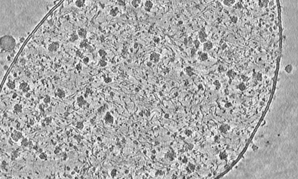
Liang Xue, a PhD student in the Mahamid Group at EMBL Heidelberg, used cryo-electron tomography to capture this detailed image of a Mycoplasma pneumoniae cell – a tiny bacterial cell used as a model system for cellular structural biology. The image beautifully depicts the cell membrane – made from a double layer of molecules known as phospholipids – and the proteins that are bound within it, as well as individual ribosomes (large dots), DNA (filamentous structures), and many other macromolecules (smaller dots) carrying out various activities.
The technology of cryo-electron microscopy, which was recognised by the 2017 Nobel Prize in Chemistry, has origins dating back to the 1970s. However, in recent years this technology, combined with modern image processing, is undergoing what has been referred to as a “resolution revolution”, making it one of the most powerful techniques for determining the structure of molecules in 3D.
By working together with leading companies in the field, EMBL provides scientists with access to the most recent available techniques and drives the evolution of new generations of microscopes, further increasing their capabilities.
Credit: Liang Xue, Julia Mahamid/EMBL
If you have a stunning picture of your science, your lab or your site, you can submit it here.


