
A better look at a human RNA polymerase
High-tech imaging illuminates the structure and function of a key enzyme in the cell, lighting the way to better therapeutics
In 2017, Julia Roberts and Owen Wilson starred in a poignant movie called Wonder, about a school-aged boy with significant facial deformities who was adjusting to life after more than 20 surgeries for a rare developmental disorder, Treacher Collins syndrome. Now, new research that captures the mutations of a mere enzyme could provide clues to several rare conditions, including Treacher Collins syndrome, characterising their molecular origins.
EMBL researchers have produced some of the most detailed data yet on an enzyme called RNA polymerase III (Pol III), which has been associated not only with rare medical conditions, but also with certain types of cancer, neurodegenerative diseases, other developmental disorders, and even a susceptibility to the varicella zoster virus, which causes chickenpox and shingles. Their findings, published in Nature Structural and Molecular Biology, illuminate the structure and function of human Pol III in different functional states.
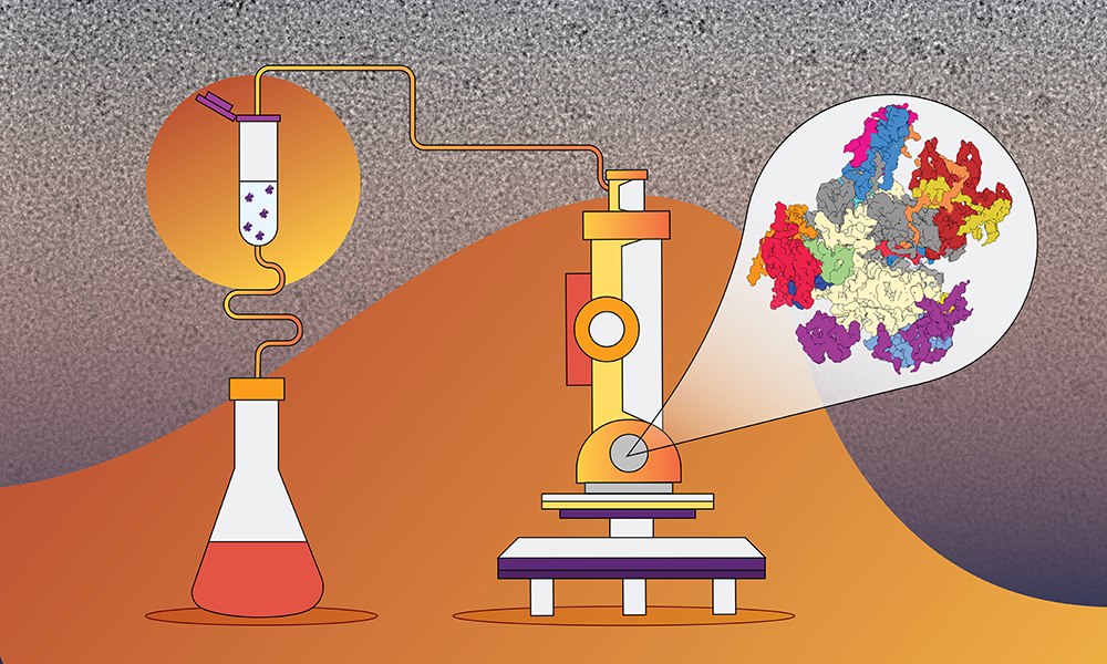
“Technology has advanced so significantly that our raw imagery with cryo-electron microscopy – although pretty fuzzy to the untrained eye – yields high-resolution data on this molecular structure, so we can observe how and to what degree mutations affect the enzyme’s function on a molecular level,” says Agata Misiaszek, one of the two lead authors and a PhD student in the Müller group at EMBL.
Human Pol III’s structure has been tremendously hard to observe using traditional techniques, such as X-ray crystallography. It has only been with the advent of modern cryo-electron microscopy (cryo-EM) and genome editing with the powerful tool known as CRISPR–Cas9 that researchers have begun making progress in understanding what Pol III looks like and how it works in human cells. In fact, the Müller group determined the Pol III structure in yeast in 2015 because of advances in cryo-EM.
In this case, the Müller group used a specialised way of culturing human embryonic kidney cells with a medium that allows for unencumbered suspension, rather than placing cells on a flat Petri dish. The group was then able to isolate Pol III from these cells and flash freeze it for the high-resolution cryo-EM analysis.
The group’s findings incorporate molecular insights about the structure of human Pol III into an understanding of how misregulation of the enzyme could be linked to human diseases. They also advance overall understanding of RNA synthesis by Pol III. In collaboration with Aleix Lafita and Alex Bateman, both computational biologists at EMBL’s European Bioinformatics Institute (EMBL-EBI) in Hinxton, UK, the team also shed light on how this human enzyme and some of its regulatory cofactors evolutionarily differ from their counterparts in other species, such as yeast.
Knowing more about human Pol III’s vulnerability to mutation helps to drive forward medical research.
“This atomic detail in imaging is still relatively new, but increasingly it allows us to get more information. In something like cancer, that means researchers could potentially target this polymerase and inhibit the disease,” says group leader and senior author Christoph Müller. “Pol III, which is one of the more abundant complexes in cells, has been implicated in cancer, ageing, and other syndromes where we can now point to specific mutations in Pol III that might be responsible.”
“What’s also exciting about the findings and this method is that it can potentially also be used for structural investigations of other complex molecular machines,” says Mathias Girbig, the paper’s other lead author and a PhD student in the Müller group. “We were able to map out about 99% of the disease-related mutations because of the incredible detail we achieved with our approach. Now this foundational work can potentially contribute to developing new drugs and more precise medical intervention.”


