NovoTags: AI-designed proteins help scientists see inside living cells
Researchers have designed synthetic and inducible proteins that bind to bright fluorescent dyes with high specificity and affinity, significantly expanding the toolkit for multicolour imaging of proteins inside cells
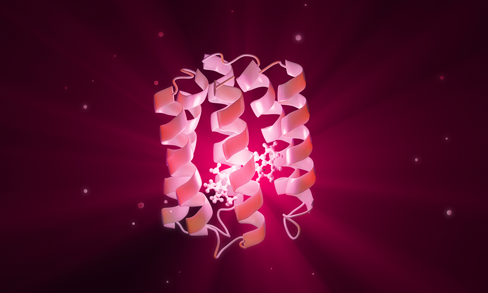
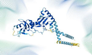
3D rendering of a NovoTag, showing the de novo designed protein around a Janelia Fluor dye.
Credit: Steffen Klein/EMBL
Summary
A team of researchers from the University of Washington, Janelia Research Campus, and EMBL Heidelberg developed a new class of fluorescent imaging tags called NovoTags.
This new tagging system is based on an AI model developed by Nobel Laureate David Baker, which allowed the scientists to create proteins from scratch that bind to exceptionally bright fluorescent dyes developed by Luke Lavis at the Janelia Research Campus.
NovoTags will allow researchers to identify specific proteins inside cells and observe many at once, accelerating the study of protein-protein interactions.
Future developments in this technology could also help researchers combine fluorescence microscopy with in situ cryo-electron tomography, providing deeper insights into structure-function relationships inside cells.
Cells are like metropolises, home to millions of molecular residents. If one were to stand atop a high-rise, trying to identify most of its inhabitants would seem an impossible task. Even with the sophisticated imaging tools currently available to scientists, it is challenging to zoom in on specific molecules and view them with a high degree of detail.
To address this limitation, researchers in the Baker Lab at the Institute for Protein Design, the Lavis Lab at Janelia Research Campus, and the Mahamid Group at EMBL Heidelberg have developed a new class of synthetic fluorescent protein tags, called NovoTags. The collaboration culminated in a paper, now published in Science, describing how versatile NovoTags can help locate specific proteins and probe their interactions inside human cells using an array of advanced light microscopy techniques, including super-resolution fluorescence light microscopy.
The project combined two complementary strengths: the AI-driven de novo protein design pioneered by David Baker, who was awarded the Nobel Prize in Chemistry in 2024 for this innovation, and the advanced microscopy technologies at EMBL, especially through the support of EMBL’s Advanced Light Microscopy Facility (ALMF).
Designing a tag from scratch
Steffen Klein, a structural biologist in EMBL’s Mahamid Group, spent six months in the Baker Lab to develop novel protein tags for fluorescence light and cryogenic electron microscopy. This research stay was made possible through the ARISE Programme – a career accelerator programme that allows researchers and engineers to advance technology development.
Baker Lab graduate student Long Tran first developed the NovoTag binders against three Janelia Fluor (JF) dyes spanning the visible spectrum, complementing existing systems such as HaloTag and SNAP-tag and expanding the possibilities for multicolour imaging. These binders are small, de novo designed proteins that can be genetically ‘tagged’ to proteins of interest and that can bind specific dyes, making them visible using fluorescence microscopy techniques.
“This study shows that designed proteins can modulate a fluorophore’s photophysical properties, offering new insight into design principles that govern photophysical function in protein-fluorophore complexes,” said Tran.
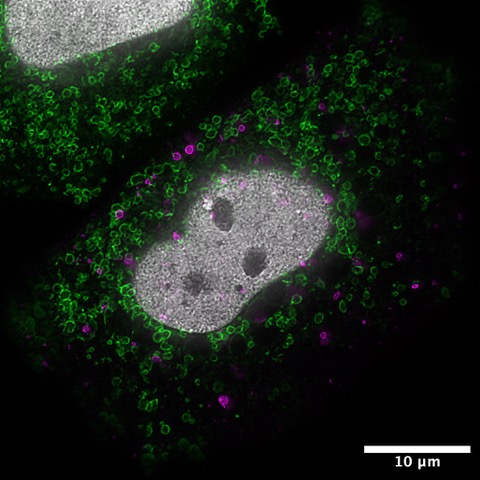
Super-resolution STED fluorescence microscopy image of a HeLa cell expressing the three NovoTags, targeting different cellular compartments: endosomes (magenta), mitochondria (green), and chromatin (white). Credit: Steffen Klein/EMBL
Klein then developed a split, inducible version, named NovoSplit, engineering a molecular switch that only assembles once its target dye is present. “A central aspect of the NovoSplit system is inducibility,” Klein pointed out. “We wanted to control protein-protein interactions, so we needed a chemically inducible system.” Essentially, this allows scientists to ensure that two proteins being studied only come in close contact with each other when an external agent – a dye – is added to the cell environment.
“Back at EMBL, I characterised the NovoTags and NovoSplit in human cell lines using advanced microscopy, particularly live STED microscopy and fluorescence lifetime imaging microscopy (FLIM),” said Klein. “By combining spectral separation with fluorescence lifetime information, we can potentially distinguish many more labels than would be possible using spectral separation alone.”
This means that in the future scientists could use this tagging system to simultaneously view or track up to 30 different proteins, by labelling each using a unique combination of emission spectrum (i.e. colour) and fluorescence lifetime (i.e. how long something emits light). The NovoTag sequences and complementary dyes are freely available to the scientific community.
The Baker and Lavis labs have already begun expanding the collection of NovoTags supported by AI@HHMI, a $500 million initiative by the Howard Hughes Medical Institute. “The combination of de novo designed proteins and synthetic fluorophores will rapidly expand the toolkit for labelling in living systems, complementing (or even supplanting) the existing approaches using fluorescent proteins and tags based on natural enzymes,” said Luke Lavis, Senior Group Leader at HHMI’s Janelia Research Campus.
Towards the next generation of cryo-CLEM labels
In essence, the NovoSplit system developed by Klein works quite simply. The NovoTag is split into two halves, each linked to a different protein in a cell. When a specific JF dye is added to the system, it acts as a molecular glue that assembles the two parts, allowing scientists to control the interaction between the two proteins.
Further exciting applications are in store. Cryo-correlative light and electron microscopy (cryo-CLEM) is a cutting-edge imaging method where the same cell can be viewed under a light and an electron microscope in a fully native state. The resulting images can be overlaid on each other to yield information neither technique could achieve alone.
Julia Mahamid’s group at EMBL, together with collaborators, has been at the forefront of pioneering these in situ cryo-ET and cryo-CLEM approaches. Their techniques have opened electron-transparent windows into cells, making it possible to visualise macromolecular assemblies at high resolution within their native, functional environment.
3D animated rendering of a NovoTag, showing the de novo designed protein around a Janelia Fluor dye. Credit: Daniela Velasco Lozano, Steffen Klein/EMBL
“Our NovoSplit system forms the basis for the development of inducible cryo-CLEM tags,” said Klein. Cryo-CLEM tags that are fluorescent and structurally distinctive enough to be identified directly inside cryo-electron tomograms would be a major leap for in-cell structural biology research.
This effort underscores the value of continued collaboration between the Mahamid and Baker groups. It also reflects how building these tags depends on a modern protein design pipeline. “We use RFdiffusion to design protein backbones and LigandMPNN to generate amino acid sequences,” explained Klein. “Candidate proteins are computationally filtered using AlphaFold and RoseTTAFold before being screened experimentally using yeast surface display, FACS, next-generation sequencing, fluorescence polarisation assays, and size-exclusion chromatography.”
“Ultimately, this work aims to expand the possibilities for identifying proteins directly within native cells by combining advanced fluorescence microscopy with cryo-electron tomography,” Mahamid explained. The possibilities such Cryo-CLEM tags could unlock are significant, enabling scientists to better study the busy alleyways of cells and their residents.
Some parts of this news article were adapted from a feature article published by HHMI, USA.