
Spotlight: Shedding light on deadly parasites
Researchers at the Kowalinski group focus on the RNA processing in Trypanosoma brucei, a parasite causative of potentially fatal tropical diseases
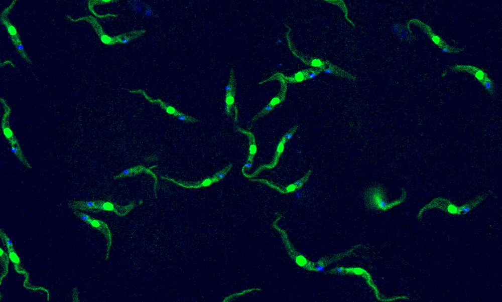
Did you know that parasites could glow green?
The image above shows a single-celled organism the size of a dust particle capable of causing deadly tropical diseases in both humans and livestock. Parasites of the Trypanosoma species are mostly found in sub-Saharan Africa, where they spread to their hosts through the bite of the tsetse fly (Glossina species). Trypanosoma brucei is also one of the few parasites known to cross the blood brain barrier, making the infection potentially fatal.
This image was taken by Luciano Dolce, postdoc in the Kowalinski lab. Here, spherical green nuclei are made visible by the use of the GFP protein – not normally present in Trypanosoma cells – that works as a fluorescent dye when inserted into the parasite’s genome. This technique is useful for observing cell morphology or localising specific proteins of interest. The blue spots show where the kinetoplast – DNA of the only mitochondrion in this cell – is located. This is seen with a fluorescent dye called Hoechst which binds to DNA, revealing its location.
The researchers at the Kowalinski group focus on how RNA processing in Trypanosoma brucei works. They study the structural basis at the molecular level – of how the proteins involved in RNA processing interact with each other, as seen in their newly published paper. They do this using a powerful and advanced microscopy technique: cryo-electron microscopy (cryo-EM). Cryo-EM involves extremely cold temperatures and an electron microscope, allowing scientists to take a look at structures thousands of times smaller than the width of a human hair. The low temperatures help with instantly freezing proteins in time, allowing the researchers to observe their shape and draw their 3D structure.
With this technique, the group was able to see how an intricate molecular machine essential to the life of the parasite coordinates RNA processing in Trypanosoma – an extraordinary glimpse of life at the atomic scale. Ultimately, with the knowledge provided by the group, researchers will be able to build better anti-parasite drugs and aid the efforts to eradicate infectious diseases.


