30 March 2023
Lab Matters, Science, Science & Technology
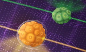
A new microscope built by EMBL researchers, based on Brillouin scattering principles, allows scientists to observe the dynamics of mechanical properties inside developing embryos in real time.
2023
lab-matterssciencescience-technology
4 January 2023
Science, Science & Technology
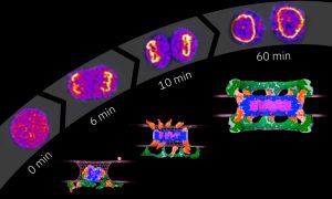
EMBL Heidelberg researchers and their collaborators reveal how the nuclear pore complex, one of the biggest molecular machines in eukaryotic cells, is assembled one protein at a time.
2023
sciencescience-technology
6 July 2021

Connections, Lab Matters
The EMBL Imaging Centre is preparing for external user access, after an on-time and on-budget build and handover to the science team.
2021
connectionslab-matters
28 April 2020
Picture of the week, Science & Technology
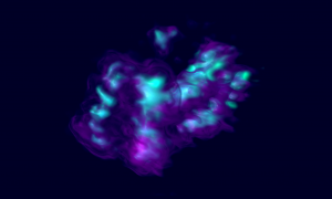
In human cells, the genetic material is packaged into 23 different DNA molecules, the chromosomes. Each chromosome is present in two copies, one inherited from the paternal sperm, and the other from the maternal egg. During most of the cell’s life, chromosomes take the shape of long,…
2020
picture-of-the-weekscience-technology
21 January 2020

EMBL Announcements, Lab Matters
Judith Reichmann will receive this year’s Paul Ehrlich and Ludwig Darmstaedter Prize for Young Researchers
2020
embl-announcementslab-matters
17 December 2019

Lab Matters
EMBL’s Jan Ellenberg reflects on the process of forming a European research infrastructure
7 November 2019
Connections, Lab Matters
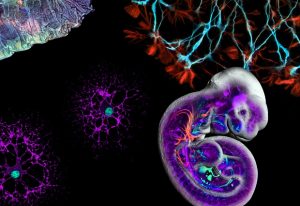
Euro-BioImaging now established as a European Research Infrastructure Consortium
2019
connectionslab-matters
10 September 2019
Picture of the week, Science & Technology
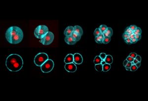
All mammalian life starts with the fusion of egg and sperm, resulting in the creation of a single cell called a zygote. This develops into an embryo through a series of cell divisions, in which the number of cells doubles at each step. Todays’ Picture of the Week was taken by Manuel Eguren of the…
2019
picture-of-the-weekscience-technology
10 September 2018
Science, Science & Technology
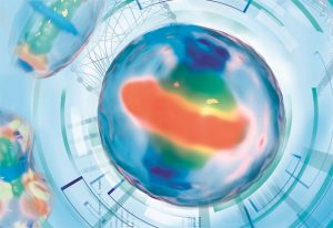
Real-time tracking of proteins during mitosis is now possible using a 4D computer model
2018
sciencescience-technology
12 July 2018
Science, Science & Technology
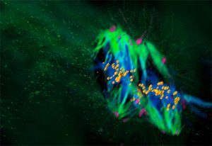
Mammalian life begins differently than we thought
2018
sciencescience-technology
7 December 2017

Science, Science & Technology
New research shows how pores form in the membrane that surrounds a cell’s nucleus
2017
sciencescience-technology
23 September 2016
Science, Science & Technology
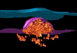
Puzzle of nuclear pore formation in growing nuclei solved
2016
sciencescience-technology
21 April 2016

Alumni, EMBL Announcements
EMBL rewards the special work of alumni through the John Kendrew and Lennart Philipson awards.
2016
alumniembl-announcements
21 April 2016

Lab Matters
EMBL scientists regularly receive prestigious awards – meet the latest honourees.
17 December 2015
Science, Science & Technology
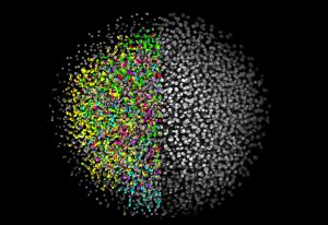
From initial development to a start-up company: Selective Plane Illumination Microscopy (SPIM) at EMBL.
2015
sciencescience-technology
14 December 2015
Science, Science & Technology
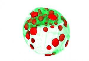
New microscope can record the first days of a mouse embryo’s life
2015
sciencescience-technology
29 September 2015

Connections, Lab Matters
Renewals and reunions: EMBL’s Nordic partners look to the future.
2015
connectionslab-matters
26 August 2015
Connections, Events
"It's like living a review!" Participants of recent super-resolution microscopy course share their highlights
24 August 2015

Alumni, People & Perspectives
EMBL rewards the special work of alumni through the John Kendrew and Lennart Philipson awards.
2015
alumnipeople-perspectives
20 August 2015
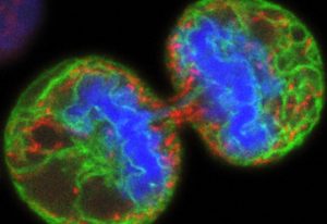
Science, Science & Technology
Collaboration between scientists reveals collaboration between lipids.
2015
sciencescience-technology
9 April 2015

Lab Matters
Major EU funding for CORBEL, facilitating access to data and biological imaging facilities.
16 March 2015
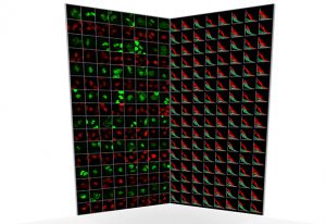
Science
New fully automated technique enables scientists to chart complex protein networks in living cells.
20 October 2014
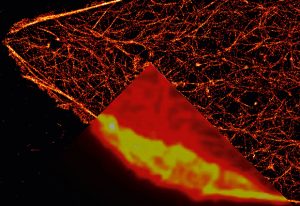
Science
How Nobel-winning work by alumnus Stefan Hell shapes and inspires current EMBL scientists' research.
17 October 2014
Science
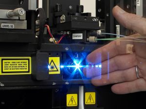
Flow cytometry: finding needles in haystacks
18 August 2011
Science
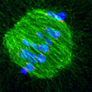
When an egg cell is being formed, the cellular machinery which separates chromosomes is extremely imprecise at fishing them out of the cell’s interior, scientists at the European Molecular Biology Laboratory (EMBL) in Heidelberg, Germany, have discovered. The unexpected degree of trial-and-error…
23 January 2011
Science
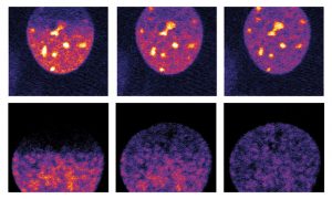
The sight of a researcher sitting at a microscope for hours, painstakingly searching for the right cells, may soon be a thing of the past, thanks to new software created by scientists at the European Molecular Biology Laboratory (EMBL) in Heidelberg, Germany. Presented today in Nature Methods, the…
2 December 2010
Science
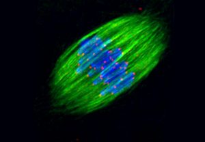
From microscopy to computer tomography (CT) scans, imaging plays an important role in biological and biomedical research, but obtaining high-quality images often requires advanced technology and expertise, and can be costly. Euro-BioImaging, a project which launches its preparatory phase today,…
1 April 2010
Science
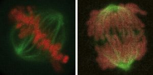
Name a human gene, and you’ll find a movie online showing you what happens to cells when it is switched off. This is the resource that researchers at the European Molecular Biology Laboratory (EMBL) in Heidelberg, Germany, and their collaborators in the Mitocheck consortium are making freely…
No matching posts found


