Intelligent microscopy
EMBL software runs experiments on its own
The sight of a researcher sitting at a microscope for hours, painstakingly searching for the right cells, may soon be a thing of the past, thanks to new software created by scientists at the European Molecular Biology Laboratory (EMBL) in Heidelberg, Germany. Presented today in Nature Methods, the novel computer programme can rapidly learn what the scientist is looking for and then takes over this laborious and time-consuming task, automatically performing complex microscopy experiments when it detects cells with interesting features.
Called Micropilot, the software brings machine learning to microscopy. It analyses low-resolution images taken by a microscope and, once it has identified a cell or structure the scientists are interested in, it automatically instructs the microscope to start the experiment. This can be as simple as recording high-resolution time-lapse videos or as complex as using lasers to interfere with fluorescently tagged proteins and recording the results.
The software is a boon to systems biology studies, as it generates more data, faster. In a mere four nights of unattended microscope operation, Micropilot detected 232 cells in two particular stages of cell division and performed a complex imaging experiment on them, whereas an experienced microscopist would have to work full-time for at least a month just to find those cells among the many thousands in the sample. With such high throughput, Micropilot can easily and quickly generate enough data to obtain statistically reliable results, allowing scientists to probe the role of hundreds of different proteins in a particular biological process.
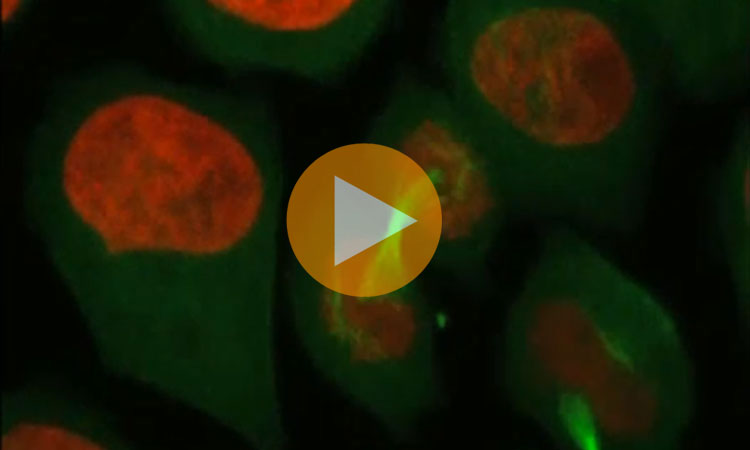
Video 1: Dividing cells automatically recorded by Micropilot.
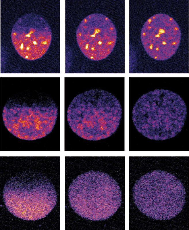
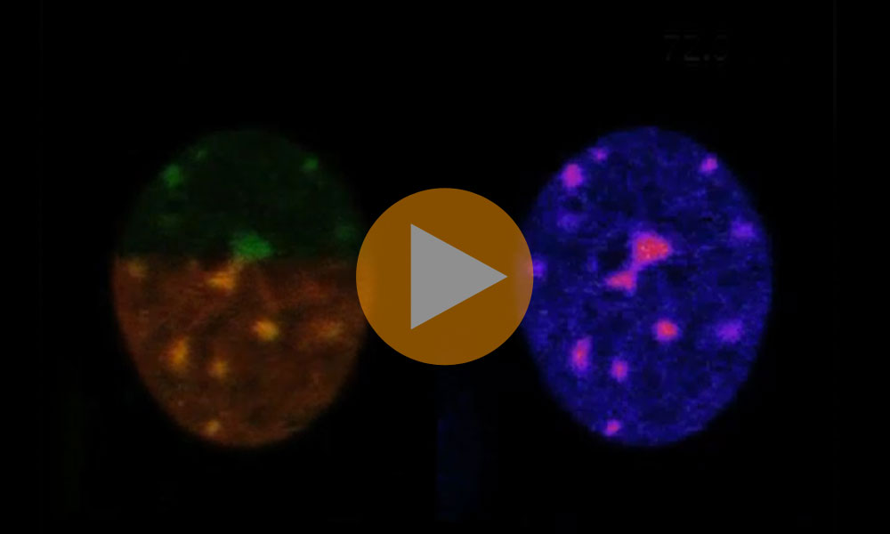
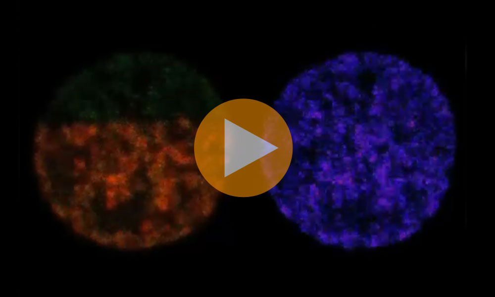
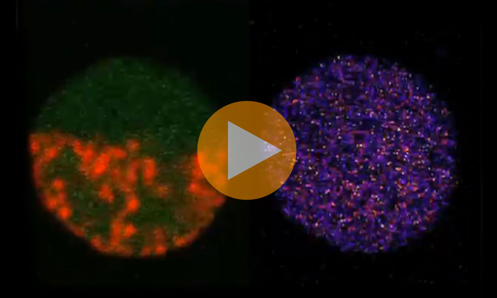
Videos 2, 3 & 4: Micropilot detected these cells and then instructed the microscope to remove fluorescent tags from proteins in half of each cell’s nucleus, and record whether fluorescently-tagged proteins moved in from elsewhere.
Video credits: EMBL
Jan Ellenberg and Rainer Pepperkok, whose teams at EMBL designed Micropilot, have used the software to deploy several different microscopy experiments, investigating various aspects of cell division. They determined when structures known as endoplasmic reticulum exit sites form, and uncovered the roles of two proteins, CBX1 and CENP-E, in condensing genetic material into tightly-wound chromosomes and in forming the spindle which helps align those chromosomes. This software will be a key tool for the European systems biology projects Mitosys and SystemsMicroscopy, for which Ellenberg and Pepperkok are developing technology.
The Micropilot software is available as open source code.