
One way crossing across the midline
EMBL researchers solve a decades-long debate on a key process for brain and embryo-development
Scientists from EMBL in Grenoble provide new insight into brain development in a paper published this week in Structure. The group from Andrew McCarthy, together with collaborators from the Grenoble-based Institute de Biologie Structurale (IBS), describe the structural changes resulting from the binding of the neural receptor Robo with the Slit protein in humans, thus providing a solution to this long-time debate.
What does this paper teach us about the human brain?
From mammals to insects, most multicellular animals share a core set of genes, conserved through evolution, that control the general structure of their bodies. Most animals have nearly-symmetrical right and left sides, and in vertebrates, each side of the body is controlled by the opposite side of the brain. For instance, the right hand is controlled by the left side of the brain: this means that some of the neuron’s long extensions, the axons, need to cross the “midline” that separates the two hemispheres of the brain.

When axons cross the midline – rich with Slit protein –, the Robo receptors on its surface bind to the protein, thus triggering a repulsive reaction that will lead the axon away from the midline and further within the brain hemisphere in which it just crossed.
Previous studies in flies and mice proved that any disruption in this process during embryo development leads to the axons crisscrossing the midline several times, and to severe malformations that will cause the death of the fetus.
What is the key finding revealed in this paper?
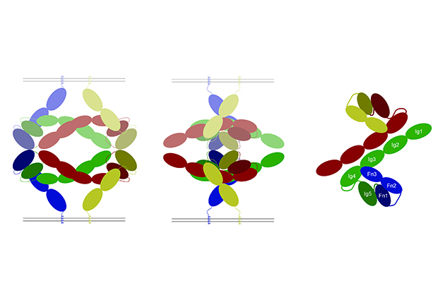
Even though scientists knew that the Robo-Slit interaction was triggering an intra-cellular reaction, the exact mechanism was open for debate: is Robo changing 3D structure or composition upon binding to Slit? Using X-ray crystallography, electron microscopy and other techniques in structural biology, we were able to prove that the composition of Robo remains intact after binding with Slit, however, its 3D structure must change. This result solves a decade-long debate and demonstrates that the signaling cascade essential for the correct guidance of the axons across the midline of the brain is triggered by a conformational change of the Robo receptor.
Could this finding have medical applications?
New studies show that there’s a correlation between the regulation of the Slit-Robo interaction and the probability of developing cancer. We suspect that when it’s over-regulated, cells can move more easily, contributing to metastasis. Unfortunately, these are complex mechanisms that we don’t really understand yet: we still have many avenues for further research….


