
Supercool structures: new cryo-EM facility
As a new cryo-EM facility is inaugurated in Grenoble, EMBL’s Michael Hons describes his role in the project
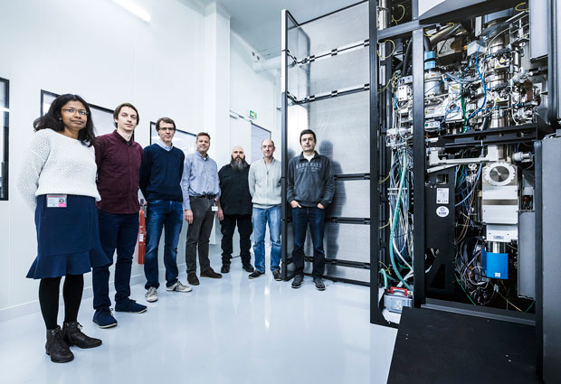
On 10 November, a major new facility for cryo-electron microscopy (cryo-EM) opened at the European Synchrotron Radiation Facility (ESRF) on the European Photon and Neutron (EPN) science campus in Grenoble. Funded largely by the ESRF, the project is run in collaboration by three organisations on the EPN campus: the ESRF, the Institut de Biologie Structurale (IBS), and EMBL. The cryo-EM facility is open to researchers from ESRF’s 22 supporting states, with access given on the basis of scientific merit. The new microscope complements the existing electron microscopy, X-ray, and neutron facilities on the EPN campus, allowing researchers to access a range of techniques for understanding the structure of biological molecules. EMBL scientist Michael Hons explains.

Tell me about your involvement in the project so far
I’m one of three scientists responsible for the day-to-day operation of the microscope, alongside Eaazhisai Kandiah from ESRF and Grégory Effantin from IBS. When I arrived here in April the place where the microscope was going to be installed was still a construction site, but it wasn’t long before the parts of the microscope were delivered and installation began. This is a major task: for many people the word ‘microscope’ might conjure up an image of something that can sit on a tabletop, but the Titan Krios microscope we are using in this facility is twice the height of a person and weighs well over a tonne. The extremely high resolution and the level of automation that can be achieved with the Titan Krios make it one of the most powerful electron microscopes currently being used in structural biology. An instrument like this is not ready to use the moment it’s unpacked, since each of the microscope’s components has to be carefully set up and aligned. It takes several weeks for construction, alignment, and testing.
I’ve also been involved in several projects using the pre-existing EM facilities on the EPN campus. There are many researchers here with intriguing questions they’d like to answer using structural biology, so it really seems like the right time to set up this new facility. It’s been a great experience to have people coming to me with a variety of samples they’d like to study using electron microscopy.
What’s special about the new facility?
It’s still rare to have a high-end cryo-EM facility which is open to a broad multinational community of scientists. From a global perspective, the time available to use the instruments at these facilities is not sufficient to meet the level of demand from scientists. A microscope like ours is beyond the budget of most universities, and most are located in labs where they are only accessible to people at the same institute or on the same campus. Our facility will attract scientists from across ESRF’s 22 supporting states.
Another important feature is the way in which the facility is being run, with scientists from ESRF, IBS, and EMBL. Our team brings together people with a diverse range of backgrounds, and with the experience needed to work collaboratively with researchers from other institutions.
Has there been a lot of interest from scientists?
Already, the level of interest is impressive. Earlier this year we had a very successful cryo-EM symposium here at the site, involving researchers from all over the world. Many people were keen to find out when the facility will be ready. It’s possible to apply for time on the microscope ranging from one up to a maximum of three days per session, so we’ll have many users each year. We believe the facility will have a very high scientific impact.
Why does structural biology appeal to you?
One thing that motivates me is the opportunity to visualise things beyond the usual range of our senses. That could be on a very large scale like in astronomy, but it can also be on a small scale like in molecular biology. Understanding the geometry of biomolecules and using that to understand their function is really fascinating. It’s a bit like seeing the mechanics of a machine for the first time, if you didn’t know what it was used for. Its purpose might not be obvious at first, but if you play around with it, see the internal parts and how they look, how they move, you can begin to get an idea of what its function is – the idea behind its construction. In structural biology we’re doing something similar: trying to understand what a biological structure is meant to do; figuring out the underlying concept.
To apply for beamtime, click here.
How cryo-EM works
Cryo-electron microscopy (cryo-EM) is a method for studying samples that involves rapidly freezing them at very low temperature and imaging them with a beam of electrons. By using electrons, much finer details can be detected than is possible with a conventional microscope, which uses visible light. The advantage of cryo-EM over other types of electron microscopy is that samples can be imaged in a state that is very close to the way they appear in nature. The very rapid freezing process prevents the formation of ice crystals, so the appearance of the sample is the same as in its unfrozen state. Freezing samples also makes it possible to avoid various types of chemical and physical preparation that are usually required in electron microscopy, which can distort the sample.
Cryo-EM is now an important tool for helping scientists determine the structures of biological molecules. The standard technique for doing this is X-ray crystallography, but that requires well-formed crystals of the molecule you want to study, which can be difficult or even impossible to obtain. For cryo-EM the sample does not need to be crystallised, and recent improvements in the technique mean that it now provides a similar level of structural detail to X-ray crystallography. Cryo-EM is therefore developing into a standard method complementing existing techniques in structural biology.


