Live from the scene: biochemistry in action
New microscope follows single molecules by the millisecond
Researchers can now watch molecules move in living cells, literally millisecond by millisecond, thanks to a new microscope developed by scientists at the European Molecular Biology Laboratory (EMBL) in Heidelberg, Germany. Published online today in Nature Biotechnology, the new technique provides insights into processes that were so far invisible.
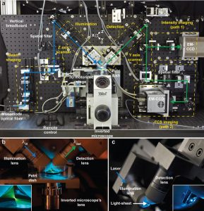
By combining light-sheet microscopy and single molecule spectroscopy, the new microscope can record the fluorescence of every pixel within view, and take snapshots at intervals of less than one millisecond. With it, scientists can watch and measure very fast processes, such as the way molecules diffuse, across a whole sample, even one containing several cells. This is a considerable step up from previous techniques, based on confocal microscopy, in which researchers could only observe at most a few isolated spots in a sample at a time.
“It’s really visual biochemistry,” says Malte Wachsmuth, who developed the microscope at EMBL. “We can follow fluorescently-tagged molecules in whole live cells, in 3D, and see how their biochemical properties, like interaction rates and binding affinities, vary throughout the cell.”
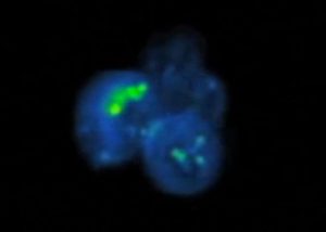
Until now, chromatin – the combination of DNA, RNA and proteins that forms chromosomes – had been observed in two states: wound tightly together, with most of its DNA inaccessible to the cell’s gene-reading machinery, in which case it is called heterochromatin; or loosely packed and easily readable, called euchromatin. But when they used the new microscope to measure the interaction between chromatin and a protein called HP1-α, the EMBL scientists made an intriguing discovery.
Credit: EMBL/H.Neves.
“In some areas that look like euchromatin, HP1-α behaves as it would in the presence of heterochromatin,” says Michael Knop, now at the University of Heidelberg, Germany. “This suggests that chromatin may also exist in an intermediate state between hetero- and euchromatin, which was not observable before in living cells.”
By providing a tool to watch molecules that move very fast, the scientists believe this new microscope will help to investigate processes ranging from the role of growth hormones in cancer to the regulation of cell division and signalling and the patterning of tissue development in the embryo.


