Choreographed origami
Folding ribosomal RNA requires paired tagging sequence
In a nutshell:
– To fold RNA that makes ribosomes, pairs of methyl tags must be added in specific order
– Detailed 3D structure of folding machinery attached to RNA
– Obtained by combination of nuclear magnetic resonance (NMR) and small angle neutron scattering (SANS)
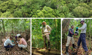
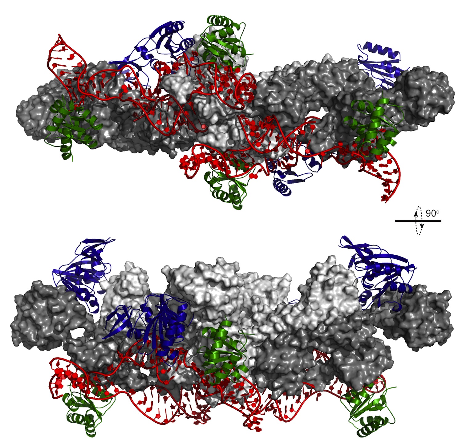
An important step in building ribosomes – the cell’s protein factories – is like a strictly choreographed dance, scientists at the European Molecular Biology Laboratory (EMBL) in Heidelberg, Germany, have discovered. To build these factories, other ‘machines’ inside the cell have to produce specific RNA molecules and fold them into the right shape, then combine the folded RNA with proteins to form a working ribosome. Like a budding origami artist pencilling in the folds, the cell uses tags called methyl groups to help mark where and how an RNA molecule should be folded. In work published online today in Nature, the scientists have discovered that pairs of these tags are added in a specific order. The study combined nuclear magnetic resonance at EMBL and neutron scattering at the Institut Laue-Langevin (ILL) in Grenoble, France.
Led by Teresa Carlomagno at EMBL, the scientists were able to determine the 3D structure of the complex that adds methyl tags to the RNA, with the RNA molecules attached. They discovered that the different components of this tagging machine pair up and move in sequence, like dancers following a set choreography.
“We found that the complex has four copies of each protein, and four methylation sites on the RNA, but those methylation sites aren’t all the same,” Carlomagno says. “They come in pairs, and one pair has to be methylated before the other.”
The fact that the pairs of tags have to be added in a particular order could be a way for the cell to control how the RNA is folded, and ultimately when and where ribosomes are formed, the scientists believe.
The study provides a detailed view of the complex in a form that’s very close to what’s found inside our cells. To obtain it, the EMBL scientists teamed up with Frank Gabel at the Institut Laue-Langevin (ILL) and the Institut de Biologie Structurale (IBS), both in Grenoble, France, to combine their expertise in nuclear magnetic resonance (NMR) with the Gabel lab’s skills in small angle neutron scattering (SANS).