From worm muscle to spinal discs
The origin of first vertebrate skeleton is likely older than previously assumed. And surprisingly, it probably evolved from muscle.
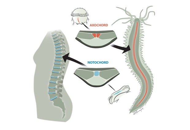
Thoughts of the family tree may not be uppermost in the mind of a person suffering from a slipped disc, but those spinal discs provide a window into our evolutionary past. Humans are part of a group of animals called chordates, whose defining feature is a rod of cartilage that runs lengthwise along the middle of their body, under their spinal chord. This structure, called the notochord, was the first vertebrate skeleton. It is present in human embryos, and is replaced with the backbone as we develop, with the cartilage reduced to those tell-tale discs. Since starfish, sea urchins and related animals have no such structure, scientists assumed the notochord had emerged in a relatively recent ancestor, after our branch of the evolutionary tree split away from the ‘starfish branch’.
People simply haven’t been looking beyond our direct relatives, but that means you could be fooled
“People simply haven’t been looking beyond our direct relatives, but that means you could be fooled, if the structure appeared earlier and that single group lost it,” says Detlev Arendt from EMBL Heidelberg, who led the research. “And in fact, when we looked at a broader range of animals, this is what we found.”
Antonella Lauri and Thibaut Brunet, both in Arendt’s lab, identified the genetic signature of the notochord – the combination of genes that have to be turned on for a healthy notochord to form. When they found that the larva of the marine worm Platynereis dumerilii has a group of cells with that same genetic signature, the scientists teamed up with alumnus Philipp Keller’s group at the Howard Hughes Medical Institute in Janelia Farm, to use state-of-the-art microscopy to follow those cells as the larva developed.
They found that the cells form a muscle – which they named the axochord – that runs along the animal’s midline, precisely where the notochord would be if the worm were a chordate.
A combination of experimental work and combing through the scientific literature revealed that most of the animal groups that sit between Platynereis and chordates on the evolutionary tree also have a similar, muscle-based structure in the same position.
The scientists reason that such a structure probably first emerged in an ancient ancestor, before all these different animal groups branched out on their separate evolutionary paths. Such a scenario would also explain why the lancelet amphioxus, a ‘primitive’ chordate, has a notochord with both cartilage and muscle. Rather than having acquired the muscle independently, amphioxus could be a living record of the transition from muscle-based midline to cartilaginous notochord.
The shift from muscle to cartilage could have come about because a stiffened central rod would make swimming more efficient, the scientists postulate.


