
Cell under attack
It’s almost a year since the coronavirus outbreak was declared a pandemic, affecting all our lives. While the virus continues its grip on the world, scientists are understanding it better and better, increasing our knowledge about it and opening up new ways to fight it.
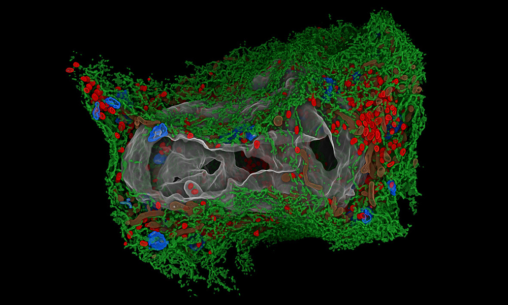
This image, taken by the Schwab team and the Electron Microscopy Core Facility at EMBL Heidelberg, in collaboration with Ralf Bartenschlager from Heidelberg University, shows a 3D rendering of an infected cell, only 24 hours after the virus started attacking it. The red spots visible in the image are a result of the virus: two membrane layers forming a balloon, within which the viral genomes are multiplied and released to be incorporated into new virus particles.
This study, one of many performed by EMBL researchers in collaboration with scientists from EMBL’s member states, will not only increase our fundamental knowledge of how SARS-CoV-2 interacts with its host, but will also help in the development of antiviral drugs.
Other colours in the image show the endoplasmic reticulum (green), Golgi apparatus (blue), cell nucleus (grey), and mitochondria (brown).
Credit: Julian Hennies, Schwab team/EMBL
If you have a stunning picture of your science, your lab or your site, you can submit it here.


