Chan Zuckerberg Initiative recognises EMBL scientists
EMBL tech trailblazers awarded for multidisciplinary collaborative projects that drive forward research
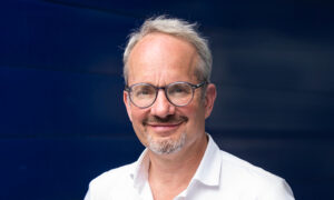
Christian Tischer leaves behind the stark white walls of his home office to bask in a sensory contrast of autumnal colours and the musty scent of decaying leaves. Even while he tries to break away so he can return to his work with fresh perspective, the daily puzzles continue to occupy his mind. Nevertheless, it’s just the right retreat to help centre his thoughts, and has become a routine part of his scientific process.
A physicist who has spent a total of 15 years at EMBL, Christian is one of four EMBL scientists recognised this week by the Chan Zuckerberg Initiative (CZI) for their expertise as technology developers in the life sciences. “CZI realised that an effective way to help science is to enable technology,” he says.
All of these EMBL recipients are bold, unconventional problem solvers. Claire Deo, Anna Kreshuk, Robert Prevedel, and Christian Tischer look for novel, elegant solutions that don’t just address one specific problem. And though none of them is a biologist, they use technology to raise the bar for biological research. It’s also clear that a pandemic may have changed the way they work, but not their drive.
“Our goal is to support the advancement of imaging technologies and provide access to and training on these state-of-the-art tools so that researchers can drive towards discoveries,” says CZI Imaging Program Officer Stephani Otte. “By collaborating closely with the imaging community and providing both funding and expertise in technology development, we hope to help make the next breakthroughs in imaging possible.”
Transforming images into something measurable
Heading EMBL’s Centre for Bioimage Analysis (CBA), Christian’s job is to support scientists in turning their microscopy data into something that can be measured, enabling them to draw quantitative and biophysically meaningful conclusions from images.
Christian’s CZI award is an Imaging Scientists grant that will fund his position for the next three years, with the possibility of an extension for a further two years. He is one of only 39 scientists worldwide to receive this honour in 2019.
At EMBL, he has worked as a Staff Scientist in the Advanced Light Microscopy Facility (ALMF) for the past 10 years, and for three years has also headed the CBA. This followed postdoctoral work at the AMOLF institute in Amsterdam and a PhD at EMBL, where he studied intracellular signalling mechanisms and cytoskeleton dynamics using advanced microscopy techniques.
“Bioimage analysis is concerned with the automated quantification of microscopy images and is a relatively recent profession,” Christian says. “Decades ago, scientists would take pictures of interesting cells and publish one picture. Today, people like me help them convert multiple images into quantitative information. At the beginning, we have images from an organism; in the end, we have numbers.”
Biologists producing data consistently outnumber the analysts developing novel algorithms and providing services in bioimage analysis. This is why CZI sees supporting these analysts as an important need for advancing science.
“This is where we bottleneck,” Christian says. “Biologists come in with their data, but very often it’s not just about straightforward image analysis. You have to really discuss with them the biological question they want to answer. I’ve had to learn a lot about biology. What makes this job both interesting and challenging is that it’s extremely interdisciplinary.”
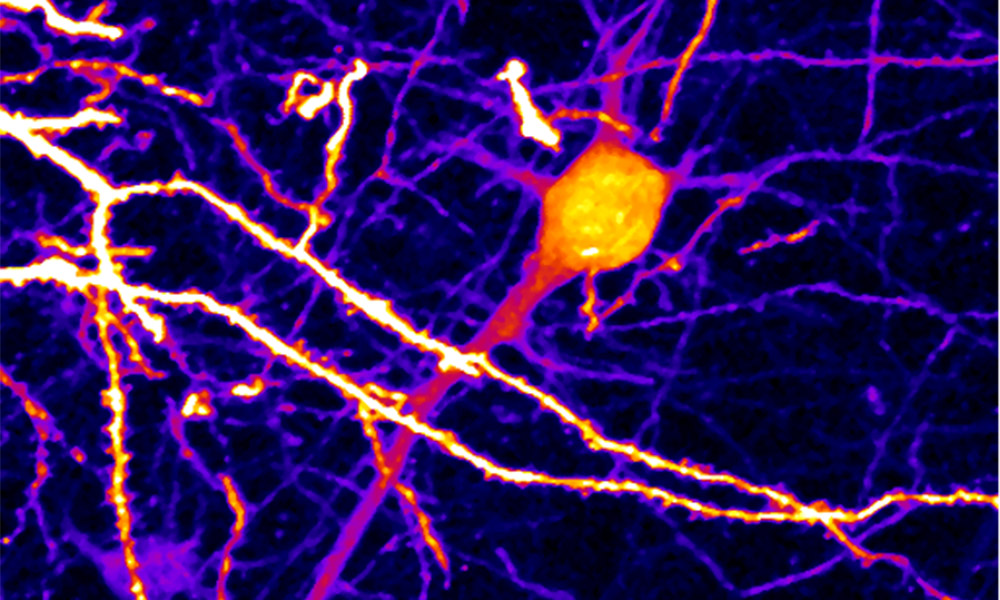
Using light and sound to see deeper into biological tissue
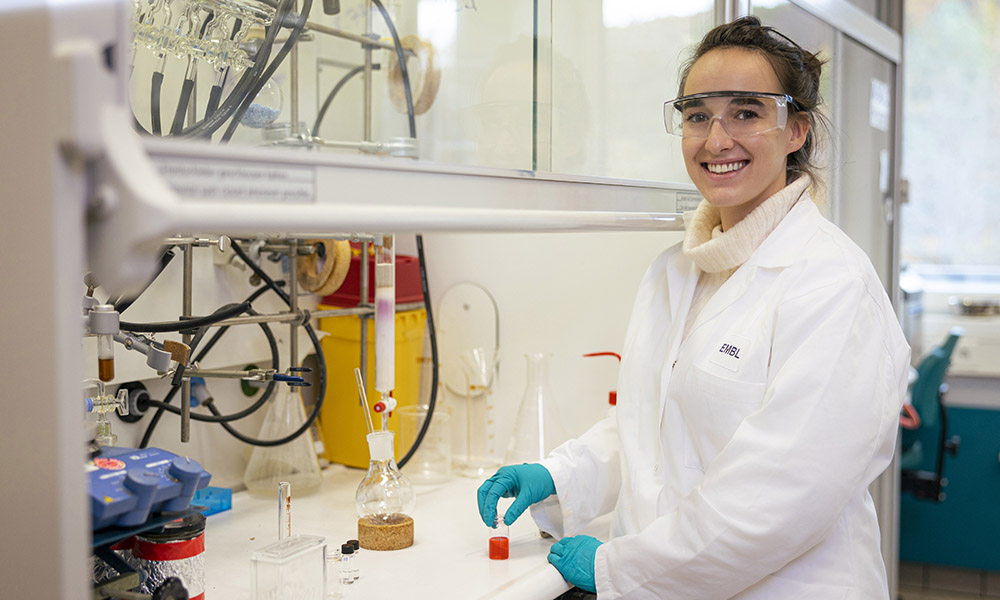
Claire Deo has a rainbow of molecular probes that will play a vital role in developing a hybrid microscope that harnesses light, sound, and adaptive optics to take non-invasive microscope imagery to a deeper level than ever before.
That’s the goal for this second CZI award, in ‘Deep Tissue Imaging’, which recognises EMBL group leaders Claire Deo and Robert Prevedel. The two EMBL scientists – one a chemist, the other a physicist and microscopy expert – will combine their talents with Sylvain Gigan, a colleague at the Kastler Brossel Laboratory at the École normale supérieure in Paris, known for his work in adaptive optics.
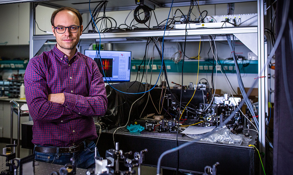
“My lab exploits photoacoustics, a technique that creates images using light and sound,” Robert explains. “Claire develops sensors or probes with molecules that can light up in either the sound or light modality, and Sylvain is an expert in a field known as ‘extreme’ adaptive optics.”
Adaptive optics is best known in astronomy, where it’s used to adjust for atmospheric distortion in telescope images. In microscopy, it can be used to adjust for the scattering of light or other signals, which is an unavoidable part of looking deeper into tissue. Light can’t penetrate biological tissue without quickly becoming diffuse, while the longer wavelength of ultrasound allows it to penetrate further, but makes it less able to resolve small details.
Adaptive optics will help to resolve these problems by seizing on the strengths of the different imaging modalities, enabling the team to extend the high-resolution field of view deeper into biological tissue. The million-dollar CZI award supports their efforts over the next two-and-a-half years.
Claire’s postdoctoral research at the Howard Hughes Medical Institute’s Janelia Research campus near Washington, DC, took her chemistry background to a new level. The researchers there were predominantly imaging cells, and Claire quickly found her niche as a developer of chemical tools in this field of microscopy. At EMBL, she uses this expertise in creating new molecular tools to advance imaging technology. “I’m currently developing molecules that turn on and off in response to light, to produce fluorescence or a photoacoustic signal,” she explains. “What really drives me is finding ways to make and apply new molecular tools to enable new scientific discoveries.”
Robert started his transition from physics to biophysics as a postdoc in Vienna, where he looked at ways to develop high-speed imaging techniques to record from neuronal circuits. He’s excited about potential neuroscience applications for the technologies he and Claire are developing. “At the moment, we can probe to depths of around 0.5 mm inside a mouse brain and observe its neuronal activity,” he says. “But we want to go deeper, to study other important brain regions such as the hippocampus, which plays an important role in memory formation and learning.”
Automating science so researchers can do more
Anna Kreshuk builds and fine-tunes computational methods to solve scientists’ research problems – sometimes before they were aware such a solution could exist.
Anna’s CZI award provides year-long funding under the heading of ‘Essential Open Source Software for Science’. It will enable her to take her easy-to-use machine learning software, ilastik, and build bridges from it to other open source tools, resulting in an improved user interface and faster execution. “It’s extremely difficult to find funding for the essential but mundane tasks that make software so much better on the inside, but are not necessarily obvious to end users,” explains Anna. “CZI provides that kind of funding, allowing us to develop better software that is more maintainable, more future proof, and more open to contributions from the community. It’s what every open source software development team dreams of!”
ilastik is a popular open source tool that brings machine learning-based image analysis to users without requiring them to have computational expertise. Anna got involved in the development of ilastik while a postdoc in the group of Fred Hamprecht, a Professor for Image Analysis and Learning at Heidelberg University. She brought ilastik to EMBL when she became a group leader here in 2018. The ilastik toolkit is a system that anyone can use to leverage machine learning algorithms that easily segment, classify, track, and count cells or analyse other experimental data. “I hope that with ilastik we will gradually move more and more image analysis tasks to the ‘easy’ category and allow people to plan their experiments as if that bottleneck didn’t exist,” Anna says.
While microscopy remains the primary application driving its development, ilastik has been used in a variety of other areas, including analysis of satellite images. Anna’s plan is to provide ilastik with an improved front end using a fast, interactive, multi-dimensional open source image viewer called napari. She will also use an open source task distribution system to speed up the software. This will reduce costs and provide an improved user experience, bringing advanced image analysis methods to a diverse range of scientists.
“For any scientist, there is an obvious thrill in thinking of something no one has thought of before, the thrill of being proven right through your experiments,” Anna says. “And for us, on top of all that, there is the joy of going beyond an intellectual exercise and seeing our work used by others for their own research.”