EMBL Imaging Centre: 2025 Highlights
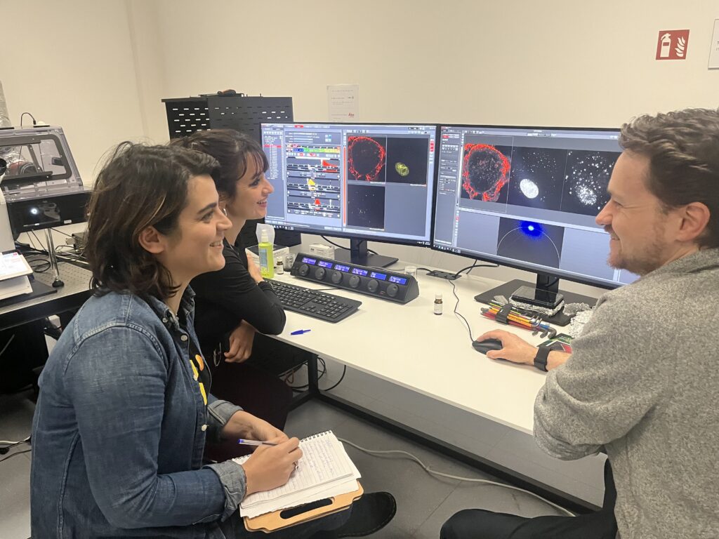
As 2025 drew to a close, we at the EMBL Imaging Centre (IC) took time to reflect on a year defined by growth, scientific progress, and technological development. It has been a rewarding year for our team, from welcoming an increasingly international community of users to advancing our in-house imaging technologies. In 2025, we supported 145 projects from 64 academic and industrial institutes, working with researchers from 18 countries, and were especially pleased to welcome users from Norway and Iceland for the first time. These milestones reflect not only the Centre’s expanding global reach, but also our shared commitment to enabling cutting-edge life science research through close support.
Advancing cancer research
A significant part of our work this year was carried out within the CanSERV initiative, providing transnational access to advanced imaging technologies for cancer research. We supported a broad range of international research teams addressing different questions in tumor biology and therapeutic development.
Among these, a project led by Prof. Lothar Dieterich used high-resolution spatial imaging to investigate interactions between lymphatic endothelial cells and immune cells within the tumor microenvironment (read more about it here). Additional projects, led by other research groups, applied advanced imaging approaches to study extracellular vesicle-mediated cancer signalling, to resolve the structure of protein complexes relevant to therapeutic targeting, and to support the development of novel antibody-based therapies.
Together, these projects highlight how state-of-the-art imaging technologies can support diverse areas of cancer research, spanning fundamental biology and translational applications.
Publication highlights
Our service efforts contributed to 16 publications this year, highlighting the broad impact of our imaging and structural biology platforms.
Among these, Wachsmuth-Melm et al. (Nature Communications, October 2025) used in situ cryo-electron tomography to visualise how Influenza A virus assembles inside cells. Their work shows how viral genome segments cluster on cellular membranes during assembly and reveals previously unseen structures of the viral matrix protein, offering new insight into how infectious virus particles are formed.
In Nature Structural & Molecular Biology (December 2025), Vidmar et al. combined cryo-EM with functional analyses to demonstrate that DNA topoisomerase I acts as a sensor of DNA supercoiling during bacterial transcription. The study provides a structural model for how transcription and DNA relaxation are coordinated to maintain proper gene expression.
A third highlight comes from Fitzpatrick et al. (Nanoscale, 2025), who tracked lipid mobility on nanoparticles at high speed and resolution using the MINFLUX technology available at the IC. The authors showed significant differences in the diffusion and mobility of lipids within silica-supported lipid bilayers, labelled with a caged dye. By tracking single fluorophores with µs and nm precision on the nanostructures, diffusive behaviours that are not accessible to other methodologies could be directly observed and measured with a suite of analysis tools specifically developed for MINFLUX tracking. Tracking was further combined with MINFLUX imaging in a separate project led by Prof. Siegfried Musser, which was featured in a news article published last year.
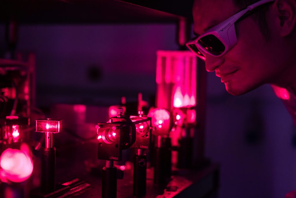
Technological innovation and development
2025 was a milestone year for technological advancement at the IC. The procurement of prestigious external funding, allowed the IC to leverage its internal engineering capabilities to spearhead new developments.
The IC is coordinating two ambitious projects funded by the Chan Zuckerberg Initiative (CZI) that aim to push the boundaries of biological imaging. Working closely with research groups across EMBL Hamburg, Rome, and Heidelberg, these initiatives will integrate advanced approaches in 3D X-ray tomography, spatial transcriptomics, and cryo-electron microscopy to establish novel workflows enabling imaging across scales, from whole tissues to individual macromolecules. Beyond advancing technology, the projects focus on developing accessible, community-driven imaging platforms and workflows that will empower the researchers to tackle complex questions in health and disease. We are grateful to CZI for their support and excited to contribute to what we hope will be a long-term collaboration advancing open and innovative imaging for the scientific community.
A second exciting milestone is the completion of two sophisticated home-built microscopes by the Centre’s engineers that expand our imaging capabilities – Oblique Plane Microscopy (OPM) and Brillouin Microscopy. OPM is a specific form of light-sheet microscopy that uses a single objective lens to illuminate an obliquely oriented plane and collect the emitted fluorescence. Light-sheet imaging can therefore be performed on an inverted microscope for samples on standard coverslips/dishes. It enables users to perform fast, gentle 3D live-cell imaging by illuminating and capturing volumetric information through a single objective lens, enabling high-resolution, dual-colour time-lapse imaging of dynamic processes in living samples with minimal phototoxicity and environmental control for physiological conditions. With the new Brillouin Microscope we bring another technology developed by the Prevedel group at EMBL into our service portfolio. It allows non-contact, label-free mechanical imaging into our portfolio, enabling researchers to quantify the stiffness and viscoelastic properties of tissues and cells at microscopic resolution. By analysing how light interacts with naturally occurring acoustic waves in a sample, this technique provides unique biomechanical insight that complements traditional fluorescence imaging and is particularly valuable for studying tissue biomechanics, disease progression, and development in living systems. Additionally equipped with a confocal scanhead it allows multimodal imaging.
Training the next generation
Knowledge transfer is at the heart of our mission, and in 2025 the IC hosted a diverse programme of training courses and job shadowing opportunities that brought advanced imaging techniques to a broad community of researchers. One of the year’s highlights was the Instruct-ERIC Hands-on Workshop on Advanced Cryo-FIB-SEM Methods, a five-day intensive course designed for participants with a fundamental knowledge in cryo-FIB, with the aim to deepen their skills in cutting-edge sample preparation and cellular structural imaging. Participants combined expert lectures with practical sessions on high-pressure freezing, cryo-CLEM workflows, automated lamella preparation, and cryo-volume imaging using our state-of-the-art instruments, while building connections with international specialists in the field.
In October, the Autumn School on Single Particle Cryo-EM offered an accessible, five-day exploration of the single-particle cryo-EM workflow from sample preparation through to 3D reconstruction. Aimed at early career researchers and facility staff new to the method, the school blended theory and hands-on learning, covering microscope operation, data processing, and best practices for high-resolution structural determination, with ample opportunity for discussion and networking.
Also in October, our Basics of Cryo-FIB Milling and In Situ Cryo-Electron Tomography course provided a foundational introduction to one of the most powerful approaches for visualising cellular architecture in its native state. Limited to just eight participants, this immersive week-long course combined theoretical background with practical training in vitrification techniques, on-grid lamella preparation, high-resolution cryo-ET data acquisition, and computational reconstruction. Lectures by leading scientists and hands-on sessions with world-class instruments helped participants build skills that they can apply to their own research projects.
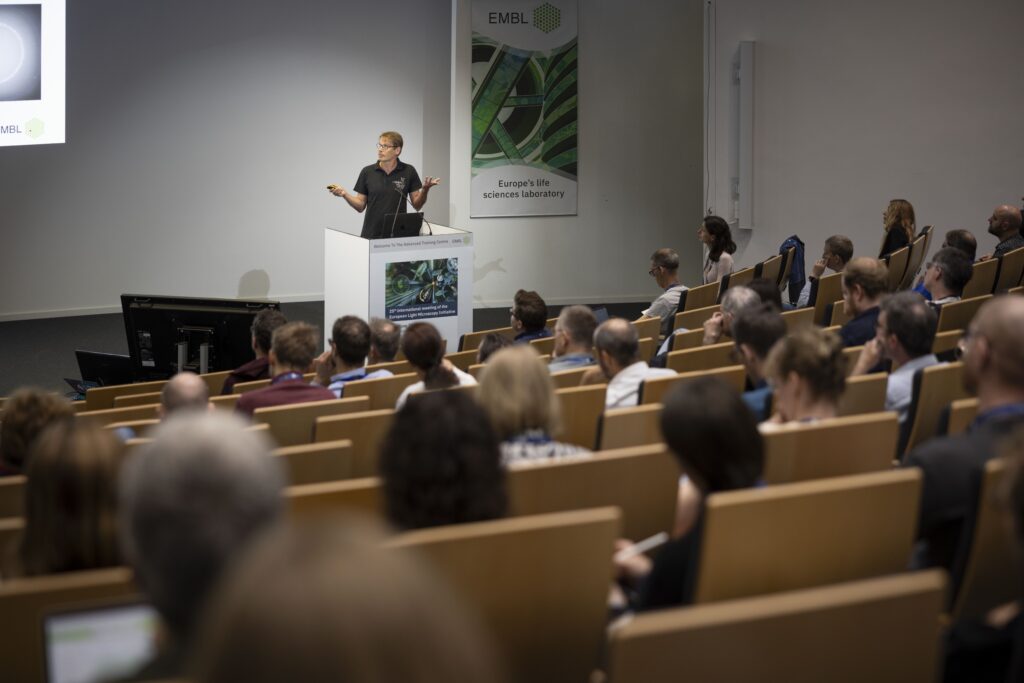
Conferences and community
The IC remained a vibrant hub for scientific exchange in 2025, both hosting and participating in key imaging conferences that brought together researchers from around the world. In June, we welcomed the 25th International Meeting of the European Light Microscopy Initiative (ELMI) at EMBL Heidelberg, the first time the event returned to EMBL since 2010. This long-standing community gathering featured a rich programme of lectures and workshops on topics ranging from probes and biosensors to super-resolution and light-sheet imaging, tissue imaging, and data analysis, alongside industry-led sessions and an extensive exhibition that facilitated lively interaction between academia and microscopy technology developers.
In October, the renowned EMBO | EMBL Symposium Seeing is Believing: Imaging the Molecular Processes of Life brought together leading developers and users of cutting-edge imaging methods to showcase how advances in microscopy are enabling real-time visualisation of biological processes across scales. The four-day meeting spanned dynamic super-resolution, intravital and tissue imaging, AI and data modelling, and emerging approaches that push the boundaries of what can be observed in living systems. With in-person and virtual participation, the symposium fostered rich discussion and collaboration across career stages and disciplines.
Looking ahead, planning is already underway for the upcoming EMBL Imaging Centre Symposium on 13 and 14 October 2026, a dedicated forum for the IC community and broader imaging field. Registrations are expected to open in Spring, and the event will further strengthen opportunities for scientific exchange, collaboration, and innovation in advanced imaging.
Moving forward: Team growth and funding
The IC’s capabilities are growing alongside its team: in July, the Boehringer Ingelheim Stiftung renewed its support to the IC with new funding, strengthening our mission to serve the scientific community. This funding will help us accelerate the development and implementation of new research-grade imaging methods, enhance access to cutting-edge technologies for the research community, and strengthen our efforts in training and support activities that broaden the reach and impact of our work. It also enables the Centre to recruit additional experts in imaging and instrumentation, ensuring that we continue to provide world-class service and innovation across scales and modalities.
As we entered into 2026, we are energised by the opportunities ahead to push the boundaries of what is visible in the life sciences, from capturing dynamic processes in living systems to revealing molecular architecture with ever finer resolution. Whether you are seeking access to our instruments, or looking for training and support, we are here to help — please reach out to ic-contact@embl.de.