
IMAGINE
New technologies to probe structure and function of biological specimens in their natural context
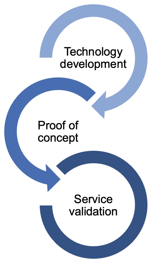
IMAGINE is large EU-wide consortium project funded by the European Union with the mission to develop the technologies needed to bridge the molecular scale of structure with the organismal scale of function and to bring those technologies not only to highly specialised central facilities and standard laboratory models, but also out in the field, at Europe’s coastline and on the ocean.