Picture Release
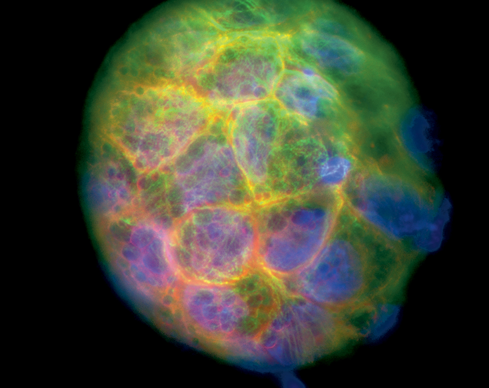
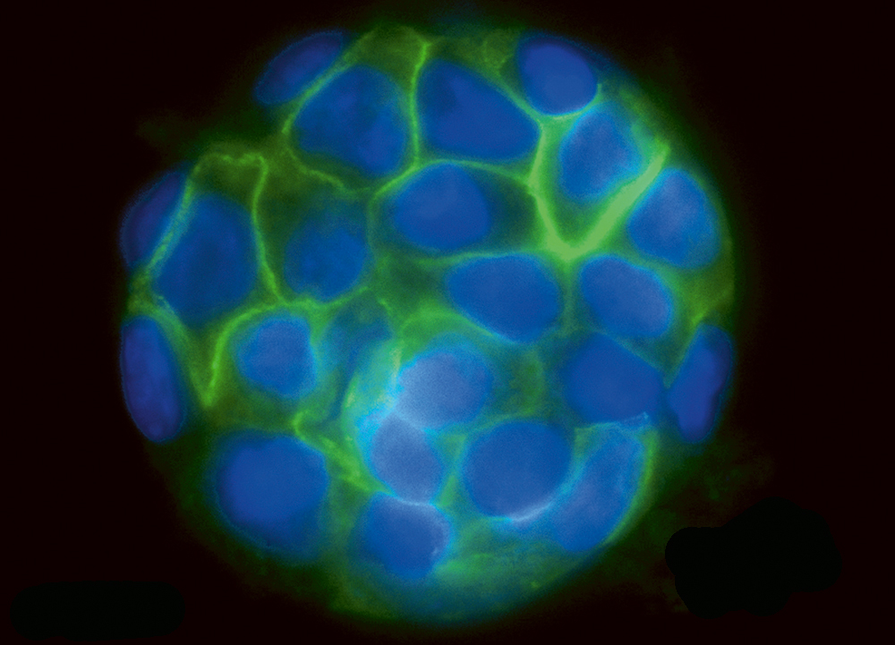
What at the first sight could be pictures of planets or other cosmic structures are actually microscope images of balls (cysts) of human kidney cells. They were taken by Emmanuel Reynaud, in the group of Ernst Stelzer at the European Molecular Biology Laboratory (EMBL), with a widefield microscope. The cysts – sacs with a liquid filled lumen surrounded by a single layer of cells – are magnified 400 times. They consist of 50 cells and have a diameter of 350 micrometers, which is five times wider than a human hair. In picture 1 (left) nuclei are stained with blue and microtubules and actin filaments, both components of the cell skeleton, in green and red respectively. Picture 2 (right) shows a similar cyst, this time with nuclei in blue and cell membranes in green.
Scientists at EMBL grow cells as cysts in three dimensional matrices to study them using a newly designed microscope, the Single Plane Illumination Microscope. Three-dimensional spheres create more life-like conditions for the cells than two-dimensional layers on flat glass slides. Cells are social creatures. They communicate with their neighbours, monitor their environment and adapt their behaviour accordingly. In flat cultures they are deprived of much of this communication. Microscope images of 3D cultures are more representative of the natural environment and show cell behaviour, shapes and arrangements as they really occur in the body.


