Drawing knowledge
A conversation about art–science collaborations and the importance of drawing in biology.
What could bring an artist and a science historian together and make them visit a research centre for molecular biology? The embryo of a fruit fly, of course.
Researchers with the academic backgrounds of Gemma Anderson and Janina Wellmann are not usually seen at EMBL. Anderson is an artist and postdoctoral research fellow. She is currently working at the University of Exeter, UK, on the collaborative project ‘Representing Biology as Process‘, together with John Dupré, a professor of philosophy of biology, and associate professor James Wakefield, a cell biologist. In her art and research, Anderson focuses on drawing as a way of generating knowledge, especially in biology. Wellmann is a historian of science and author of The Form of Becoming: Embryology and the Epistemology of Rhythm, 1760–1830. In this book, Wellmann describes how scientific observation is limited when confronted with living, moving systems and how creative decisions influence the scientific process. Both Anderson and Wellmann are fascinated by shape changes and dynamic processes in biology. “Our common interest is the question: how do you depict changes over time and how do the processes of visualising and making it intelligible co-evolve?” says Wellmann. Anderson and Wellmann want to find a way to depict dynamic biological processes through drawing, to include time and change in a two-dimensional image; a challenge that overlaps the worlds of science, art and philosophy.


During a conference in 2017, Anderson attended a talk by EMBL group leader Maria Leptin on embryogenesis and gastrulation – the process during which the embryo folds inward, transforming from a hollow ball of cells to a multilayered structure. Anderson was fascinated by the Leptin group’s research on cell shape transformation in fruit fly embryos. After the conference, she contacted Maria Leptin to talk about her work at EMBL and asked if she could visit to see for herself. When Wellmann heard about the Leptin group’s work, she decided to join Anderson on her visit.
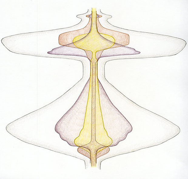
During their week at EMBL, Anderson and Wellmann observed the work of the research group in the laboratory. While Anderson gathered ideas and information so she could start drawing, Wellmann was particularly interested in the technology used to visualise the development of the fruit fly embryo. Anderson and Wellmann also spoke to scientists individually about the processes they were studying. “In the lab, they image the three-dimensional body of the embryo,” says Anderson. “But, just like with the map of the world, they then do a cartographic projection as a 2D image and flatten it, so you see the whole surface.” She started working on some first sketches based on these conversations, facing the challenge of drawing a part of the process that could not be properly visualised with the microscope. “In the process of gastrulation, part of the surface goes inside and then you don’t see it any more. So, in their images, you just get this line where that’s happening. I’ve been trying to make a drawing that actually includes that inside surface as well. It is part of the same surface, so theoretically you could flatten it all out.”

An important aspect of Anderson’s view of art–science collaboration is that drawing should not be understood as a way of merely illustrating or decorating scientific thought. Wellmann shares the same opinion. “It’s not that you use drawing as an illustration of something you have thought of before,” she explains. “Your thoughts about it evolve together with the ability to represent it.” Both agree that if drawing would be recognised and used as a valid tool for knowledge generation, this could significantly benefit scientific work. Anderson sees many reasons to include regular drawing sessions in research activities. “Scientists often say that they don’t find enough time to think,” she says. “I would say that if you carved out space for drawing in scientific practice, that also creates time for thinking.” Drawing can also create a personal connection between a scientist and the object of study, which is missing when the image is taken by a scientific instrument. “If you try to draw something that you’re working on, what you decide to keep and what you decide to leave out are quite intellectual decisions,” says Anderson. “Drawing also shows you what you understand, and you can then show that understanding to others.” This is why, during her visit, Anderson actively encouraged scientists to explain their research to her through drawing.
Sourabh Bhide, a postdoc in the Leptin group, says: “Between the first day of their visit and the last, my opinions shifted a lot. In the beginning I thought: ‘I really don’t know how this is going to help.’ But, in the end, I realised that it made me think so much about the process when I was actually trying to put it down on a single sheet of paper. It can definitely have an impact. It can give you one more dimension to your thinking that you usually miss out completely.”
This explanatory quality of drawing is what makes it such a useful tool during interdisciplinary encounters. Where language is not enough, because it is encrypted in the jargon of individual research fields, visual communication can help to break that code. During their visit, Anderson and Wellmann talked to EMBL group leader Robert Prevedel, who works on developing new microscope technologies. This brings him together with biologists, physicists and computer scientists, all of whom have to somehow explain their ideas and needs to each other. “They use basically everything that they have at hand,” explains Wellmann. “So, they do drawings, they doodle, they try to clarify the concepts when they talk about things. You’re dependent on a mixture of all kinds of representations and ways of making your ideas intelligible in words, in drawings, movements and gestures.”
Another observation made during the conversation with Prevedel was that biologists draw very differently from physicists when they’re explaining something. These differences between disciplines also apply to the perception of images: during their visit to the laboratories, Anderson and Wellmann followed the preparation and use of the SPIM microscope – based on a technology developed at EMBL that uses a thin sheet of light to illuminate only one plane of a sample at a time, minimising damage to the sample caused by light. After looking at images from the SPIM microscope, Wellmann noticed that scientists had very different opinions on how to define what they were seeing. While some referred to the images as data, others saw them as a model. “We had to figure out during the conversation that, when we were talking about images, we meant different things,” she explains.
Drawing used to be the preferred visual tool for biologists. Nowadays, it has largely been replaced by modern tools with increasing technological complexity, ranging from photography to three-dimensional data visualisation. Wellmann is fascinated by the philosophical issues that are opened up by these new imaging technologies. As an example, she describes the elimination of the ‘observer’ from the scientific process. Visual information is now captured by machines using specialised software, and only the result of this machine-driven observation is then interpreted by scientists. Anderson has questions on this topic as well: “If you’re not doing optical observation, and it’s basically a machine translating data into pixels, then what do we mean when we say that we see something, and who is seeing? I find this human–computer mediation really interesting.”

Wellmann explains that technology has a strong influence on how people think, something that can be observed in the use of new imaging methods. “The question is: how much in these visuals is framed by technology and computer programs and how much is framed by the creative choices of the scientist?” she says. “The choice of colours they use, is that a lab convention or is it something that you think about? It’s in the little details that you don’t necessarily notice. When you look at it again and again, you see that there are a lot of choices made in those depictions. Which of those are part of the scientific work, or of the imagination of the scientist? Is it just that they’re given by the technology you have at hand or that your lab uses, which is why you use them as well?”
Anderson believes that a way to actively reflect on these creative decisions during research is by drawing. Considering all the imaging technology that modern laboratories use, it seems easy to neglect drawing as an old and inaccurate method. Although drawing is still a part of biological education, the importance that is given to it appears to be decreasing. “In my experience, at the PI level I find more people drawing; at the postdoc and PhD level, not so much,” says Anderson. She thinks that there are various reasons why it has fallen out of scientific education and practice: scientists simply may not see the benefit of drawing, or may be unable to make space for it in an already packed schedule.
So, how could scientists find more time for drawing in their research? “First, you have to take drawing seriously,” says Anderson. “Then defend it in your group, say: this is part of my practice, part of how I do my science.” Anderson also observes that many scientists are often shy to start drawing if they feel they are not good at it, but this quickly changes once they start practising, and can even become difficult to stop. Emphasising the relevance of drawing during education could be a very efficient way to promote its further use in the laboratory. “Scientists, especially those involved in education, should open a space for drawing,” says Anderson.
Anderson and Wellmann both seem very inspired by their week at EMBL. “What is fascinating is to have the chance to be exposed to an environment that is unusual to me,” says Wellmann. “I have to be attentive, to be observational and really look and see and try to understand.” She observes that, to scientists, this sort of interdisciplinary interaction is unusual as well, but is very well received.
Explaining his work through drawings to someone without a biological background has been an interesting experience for Bhide. “Usually we only interact with other scientists,” he says. Bhide has promised Anderson that he will continue drawing in the future, and will report back on how this has changed his understanding of his work.
Anderson’s visit to EMBL and the project ‘Representing Biology as Process’ (2017–2020) are funded by the Arts and Humanities Research Council. Further information on art–science–philosophy collaborations can be found on the website to Andersons’ project and a blog post on the EMBL visit.