Atomic proportions
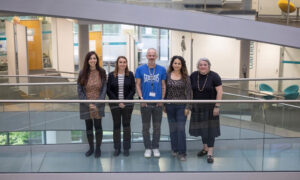
How EMBL scientists and mechanics are working together with ESRF colleagues on an upgrade for one of Europe’s big X-ray sources
Just as doctors use X-rays to study the hairline fracture of a bone in the hospital, scientists also use X-rays to explore structures that are invisible to the naked eye. But whereas the hospital’s X-ray machine can only reveal details of the skeleton beneath our skin, the scientists’ X-rays – which are produced by stadium-sized circular machines called synchrotrons – are able to delve far deeper and reveal minute details of the atomic world. They are enabling unprecedented insights into such worlds as the surface inaccuracies of metal, to the delicate fossils trapped in rocks, to the paintings hidden by their creator under famous masterpieces, to the molecular structures of proteins.
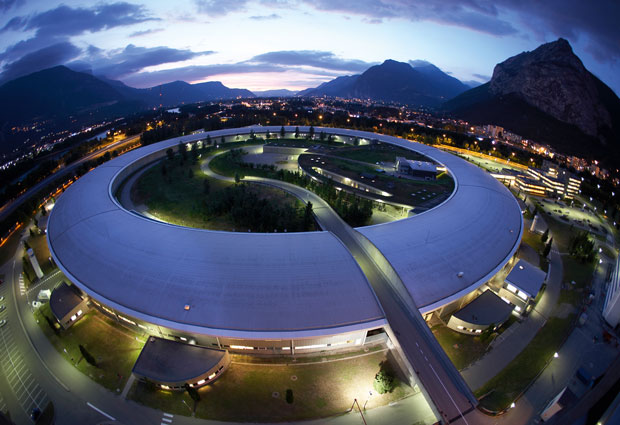
The European Synchrotron Radiation Facility (ESRF), situated at the foot of the French Alps on the European Photon and Neutron (EPN) campus in Grenoble, has been producing X-rays for scientific research for more than 20 years. EMBL’s Grenoble site also forms part of the campus. Scientists there use this synchrotron radiation to explore the 3D atomic structure of biological molecules such as proteins and nucleic acids, shedding light on how we might prevent them causing disease or harness their unique qualities for new biotechnologies. As part of a collaboration that has been ongoing for over two decades, EMBL scientists work alongside ESRF colleagues to design and operate measuring stations – or beamlines – for structural biology experiments on the 844m synchrotron ring. Now, in an extensive modernisation programme structural biologists will be able to develop techniques such as time-resolved crystallography and serial crystallography to explore, in unprecedented detail, the molecules that make up our world.
An air of excitement
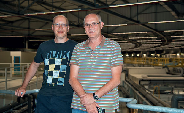
“There is definitely an air of excitement here at the moment,” says Gordon Leonard, the Head of the Structural Biology Group at the ESRF. EMBL group leader Andrew McCarthy, who works closely with Leonard as part of the ESRF-EMBL Joint Structural Biology Group (JSBG) to improve the accuracy and speed of structural biology experiments, agrees. “Our users will be able to use the traditional crystallography set-ups they are used to, as well as being able to choose from a wider range of techniques such as serial and time-resolved crystallography and even being able to take high-resolution images of individual proteins inside the cell.”
To study the structure of a protein using X-ray crystallography, scientists first need their protein samples to take on a crystalline form – much like the salt crystals produced from salt water. When put into the X-ray beam, the X-rays diffract off the atoms in the protein crystal, producing a distinct pattern that can be used to understand the molecular structure of the protein. But as scientists seek to explore the structure of larger and more complex proteins – and even multiple protein molecules bound together – the crystals that these samples produce are, conversely, smaller and more fragile.
Traditionally, scientists remove protein crystals from the plates in which they grow before taking them to the beamlines. Many proteins, such as some membrane proteins, cannot be encouraged into forming large crystals but will often form very tiny crystals. Until recently these very fragile crystals were almost impossible to extract from where they were grown. To get around this problem, EMBL team leaders Florent Cipriani and Jose Marquez developed CrystalDirect™. “This technology allows you to automatically mount individual or multiple crystals from plates and subsequently move them directly into the X-ray beam for measurements,” explains McCarthy. “We will implement this technology on the beamline for serial crystallography techniques, which will be a great help to our users.”
Extreme brilliance
To be able to measure these tiny crystals, a smaller and more focused X-ray beam is also needed. “With bigger, less compact beams, most of the X-rays will pass by such a small crystal without touching it – that is a lot of lost energy,” explains Leonard. “And the longer the crystal is exposed, the higher the risk of damage.”
The 150 million euro ESRF Extremely Brilliant Source (ESRF-EBS) project aims to address this problem. “We will produce X-ray beams up to 100 times more brilliant and focused than is currently possible,” says Leonard enthusiastically. “That’s like the difference between trying to read a book by the light of a candle in the next room and holding a powerful head torch directly over the page.”
It is opening our eyes even wider to an incredible molecular universe
This ambitious project entails replacing the current synchrotron storage ring with an upgraded version within the existing ESRF infrastructure, all the while recycling as many components as possible. “The old infrastructure will be re-used – this keeps costs and construction time to a minimum and allows us to keep the existing experimental stations fed by the storage ring,” explains Leonard.
Key to the new design are 1000 custom made, high-performance magnets. They will be slotted precisely together in a lattice designed to reduce energy loss, while guiding electrons around the ring and producing some of the world’s most compact and minute X-ray beams. To minimise disruption, much of this work will initially take place in specially constructed temporary buildings. Installation and commissioning is scheduled to begin in late 2018. “It’s a fast turnaround, but we hope we can take the first users again in spring 2020,” says McCarthy.
Smaller and smaller
But even before the ring upgrade begins, several of the beamlines themselves are being overhauled. One of these is ID23-2, the world’s first microfocus beamline dedicated to macromolecular crystallography. “It has contributed nearly 900 depositions in the Protein Data Bank (PDB), including several relating to the structures of medically important membrane proteins, but it is now over 10 years old,” McCarthy explains. “The optical configurations will get a major overhaul in order to provide an extremely high intensity beam with a micron-sized diameter. This also requires new high-precision instruments designed by Cipriani and his team for measurements, as well as new algorithms for analysing the data.”
A bright future
Meanwhile, the JSBG are planning even further into the future. “With such an intense beam, it will be possible to do time-resolved experiments,” says McCarthy. “More information will be captured in shorter time frames: we can start to ‘watch’ protein structural changes in real time by capturing multiple snapshots and putting them together to form a movie, opening our eyes even wider to an incredible molecular universe,” he explains. “The future is very bright!”