
Foetus, or placenta?
New research in Cell shows subtle differences between seemingly identical cells at a very early stage of development
When exactly does an embryonic cell decide whether it will become part of the foetus, or part of the placenta? Scientists at the University of Cambridge and EMBL-EBI shed light on this important question by studying the development of mice embryos only four cells in size. The findings, published in Cell, have implications for the understanding of mammalian development.
When an embryo first starts to develop, genetic factors influence whether its cells will become part of the supporting structure around a new organism, or part of the organism itself. Today’s study shows that seemingly identical cells in a two-day-old mouse embryo already begin to display subtle differences.
Once an egg has been fertilised by a sperm, it divides several times to become a free-floating ball of stem cells. At first, these cells are ‘totipotent’, able to divide and give rise into any type of cell, placental or organismal. Some of those cells then switch to a ‘pluripotent’ state, in which their development is restricted to generating the cells of the whole body, rather than the placenta. Understanding that switching mechanism, and when it occurs, is the subject of intense research.
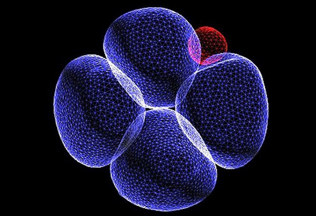
“We know that life starts when a sperm fertilises an egg, but we’re interested in when the important decisions that determine our future development occur,” says Magdalena Zernicka-Goetz from the Department of Physiology, Development and Neuroscience at the University of Cambridge. “We now know that even as early as the four-cell stage – just two days after fertilisation – the mouse embryo is being guided in a particular direction and its cells are no longer identical.”
The team used the latest sequencing technologies to understand embryo development in mice, looking at the activity of individual genes at a single-cell level. They showed that some genes in each of the four cells behaved differently. The activity of several genes in particular differed the most between cells. These genes form part of the ‘pluripotency network’: they are targeted by key pluripotency factors, including Sox2 and Oct4. The team showed that when activity of such genes is reduced, the activity of a master regulator, which directs cells to develop into the placenta, was increased.
John Marioni of EMBL-EBI, the Wellcome Trust Sanger Institute and CRUK-CI, adds: “We can make use of powerful sequencing tools to deepen our understanding of the molecular mechanisms that drive development in individual cells. Because of these high-resolution techniques, we are now able to see the genetic and epigenetic signatures that indicate the direction in which early embryonic cells will tend to travel.”
The research was funded by the Wellcome Trust, the European Molecular Biology Laboratory and Cancer Research UK.
This post was originally published on EMBL-EBI News.


