
Destined for the brain
Despite their importance, the way microglia develop and end up in the brain has remained unclear. Now, new research led by Francesca Peri, Group Leader in the Developmental Biology Unit at EMBL Heidelberg has provided new insights into this process, as described in Cell Reports.
When we think about the brain, neurons often take the lead role. Yet more than half the brain is made up of so-called glial cells that pack out the brain and help keep neurons in place, provide them with nutrients, and insulate them from each other. One type of glia, microglia, are specialised macrophages – immune cells that swallow up and destroy bacteria, viruses, cellular debris and even cancer cells – and provide the first line of defence against pathogens in the central nervous system.
Prior to the work led by Peri, microglia were known to develop from macrophage cells during the early stages of embryonic development, and it’s been suggested that any macrophage that ends up in the brain can become a microglial cell. “Some recent studies have argued that embryonic macrophages are all the same and simply adapt to their local environment,” says Peri.
Some recent studies have argued that embryonic macrophages are all the same and simply adapt to their local environment
To put this claim to the test, Peri and colleagues studied the development of microglia in the tiny zebrafish, a commonly used animal model in developmental biology. Earlier studies had shown that in zebrafish macrophages develop in two waves, and in distinct parts of the embryo. The first wave occurs 20–26 hours after fertilisation in the anterior lateral plat mesoderm (ALPM), with the second beginning 30 hours post-fertilisation in the intermediate cell mass (ICM).
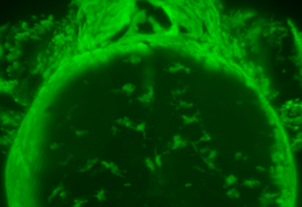
To find out where microglia come from – the ALPM or ICM – Peri and colleagues genetically engineered zebrafish so that all their macrophages expressed a protein called Kaede, which causes the cells to glow green. Kaede, however, can be made to glow red by activating it with a blast of UV light. So the team gave the cells in the ALPM a dose of UV, while in separate experiments they shone light on the ICM, and then looked to see whether any red-glowing microglia ended up in the brain. They found that only red-glowing cells from the ALPM ended up as microglia in the brain.
Peri and colleagues also identified a mutant zebrafish that lacks microglia, and discovered that the problem was a faulty version of the slc7a7 gene. “We found that in zebrafish, this gene is expressed in only a subset of macrophages in the ALPM during early embryonic development,” says Peri. And contrary to the idea that any precursors can turn into microglia, it’s only this subset that migrate to the brain to mature into microglia. “Not all embryonic macrophages are the same, and only those expressing slc7a7 are destined to become microglia,” says Peri.
Not all embryonic macrophages are the same, and only those expressing slc7a7 are destined to become microglia
To confirm the role of slc7a7 in microglia development, the team inhibited the expression of this gene in otherwise normal zebrafish, which created embryos lacking microglia similar to the slc7a7 mutants. Likewise, adding a functional copy of slc7a7 to the mutants restored the microglial population. “Our work shows that Slc7a7 is essential for getting microglial precursors in to the brain,” says Peri.
In future work Peri and colleagues hope to find out just how Slc7a7 determines the fate of macrophage precursors, and what signals these cells use to guide them to their home in the developing brain. These insights could one day provide a basis for developing therapies that selectively recruit macrophages to the brain to tackle diseases such as cancer.


